Ricardo Maliba's research while affiliated with Montreal Heart Institute and other places

What is this page?


This page lists the scientific contributions of an author, who either does not have a ResearchGate profile, or has not yet added these contributions to their profile.

It was automatically created by ResearchGate to create a record of this author's body of work. We create such pages to advance our goal of creating and maintaining the most comprehensive scientific repository possible. In doing so, we process publicly available (personal) data relating to the author as a member of the scientific community.

If you're a ResearchGate member, you can follow this page to keep up with this author's work.

If you are this author, and you don't want us to display this page anymore, please let us know.

Publications (10)


Fig. 1. Ang1 and Ang2 activate Tie2 in a time-dependent manner. Confluent BAEC were treated with Ang1 (A) or Ang2 (B) from 5 min to 15 min. Cell lysates (500 ␮ g) were prepared and immunoprecipitated with rabbit polyclonal anti-mouse Tie2 IgG. Following resolution by SDS-PAGE and transfer, PVDF membranes were probed with mouse monoclonal antiphosphotyrosine IgG, and immunoreactive bands were visualized by chemilu- minesence. Membranes were subsequently stripped using ReBlot Plus Strong stripping solution, and Tie2 protein expression was determined by Western blot analysis following incubation with rabbit polyclonal anti- mouse Tie2 IgG. Relative Tie2 phosphorylation (pTie2) over Tie2 total protein expression under various experimental conditions was calculated by normalizing DPBS control OD values to 1 (C). 
Fig. 2. Ang1 and Ang2 induce endothelial P-selectin translocation in a time- and concentration-dependent manner. Confluent BAEC were treated with Ang1 or Ang2, and P-selectin translocation was measured by cell surface ELISA. Solution buffer (DPBS) was used as control, and the basal levels of P-selectin translocation were normalized to 1. Maximal P-selectin translocation (T) was observed at 7.5 min (A) at a concentration of 1 nM (B). Similarly, Ang2-mediated endothelial P-selectin translocation was maximal at 7.5 min (C) and at 1 nM (D). As positive control, BAEC were stimulated with VEGF-A 165 (1 nM; 7.5 min). Data are expressed as relative absorbance measured at 450 nm. Values are means Ϯ SEM of P-selectin translocation obtained from at least three independent experiments. 
Fig. 3. Ang1 and Ang2 mediate endothelial P-selectin translocation. Confluent BAEC were stimulated with DPBS, PMA (1 ␮ M), VEGF-A 165 , Ang1, or Ang2 (1 nM) for 7.5 min. Cells were fixed, labeled with antisera, and visualized by confocal microscopy. Cell surface membranes were labeled with WGA conjugated to Alexa 488 and appear in green. P-selectin distribution was detected by using rabbit polyclonal anti-human P-selectin IgG followed by incubation with a secondary antibody conjugated to Alexa 555 IgG and appears in red. The presence of P-selectin within the cell surface membrane appears in yellow. Front view transparent projections (A1–F1) were made using four slices (0.64 ␮ m thick) from respective Z stacks. A 2 ␮ m-wide rectangular volume from the Z stacks was extracted to produce transverse transparent projections (A2–F2), executed using the LSM 510 software. Three-dimensional (3D) representation of the cells (A3–F3) was performed by using the SFP software; projections were created using all the slices of the Z stacks. Original magnification, 63 ϫ ; digital zoom, 2 ϫ ; bars, 5 ␮ m. 
Fig. 4. Endothelial distribution of Ang2 upon stimulation with VEGF-A 165 
Fig. 5. Endothelial P-selectin translocation mediated by Ang1 or Ang2 requires PLC- ␥ and PKC signal transduction. (A) Confluent BAEC were pretreated with selective pharmacological inhibitors for PLC- ␥ (U73122; 10 ␮ M) and PKC (Calphostin C; 100 nM) for 15 min prior to 7.5 min of stimulation with Ang1 or Ang2 (1 nM). BAEC were also stimulated with VEGF-A 165 (1 nM) 

+2

Angiopoietin-mediated endothelial P-selectin translocation: Cell signaling mechanisms
  • Article
  • Full-text available

March 2008

·

229 Reads

·

26 Citations

Journal of Leukocyte Biology

Ricardo Maliba

·

Alexandre Brkovic

·

·

[...]

·

Recently identified, angiopoietin-1 (Ang1) and -2 (Ang2) bind to the tyrosine kinase receptor Tie2 and contribute to orchestrate blood vessel formation during angiogenesis. Ang1 mediates vessel maturation and integrity by favoring the recruitment of pericytes and smooth muscle cells. Ang2, initially identified as a Tie2 antagonist, may under certain circumstances, induce Tie2 phosphorylation and biological activities. As inflammation exists in a mutually dependent association with angiogenesis, we sought to determine if Ang1 and/or Ang2 could modulate proinflammatory activities, namely P-selectin translocation, in bovine aortic endothelial cells (EC) and dissect the mechanisms implicated. P-selectin, an adhesion molecule found in the Weibel-Palade bodies of EC, is translocated rapidly to the cell surface upon EC activation during inflammatory processes. Herein, we report that Ang1 and Ang2 (1 nM) are capable of mediating a rapid Tie2 phosphorylation as well as a rapid and transient endothelial P-selectin translocation maximal within 7.5 min (125% and 100% increase, respectively, over control values). In addition, we demonstrate for the first time that angiopoietin-mediated endothelial P-selectin translocation is calcium-dependent and regulated through phospholipase C-gamma activation.

Download
Share


HIF2α reduces growth rate but promotes angiogenesis in a mouse model of neuroblastoma

February 2007

·

168 Reads

·

36 Citations

BMC Cancer

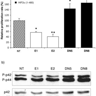
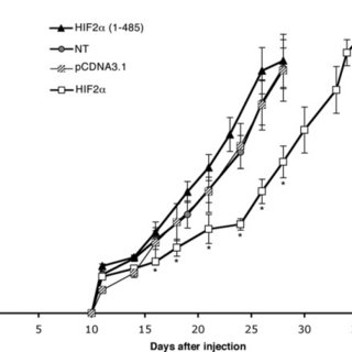
HIF2alpha/EPAS1 is a hypoxia-inducible transcription factor involved in catecholamine homeostasis, vascular remodelling, physiological angiogenesis and adipogenesis. It is overexpressed in many cancerous tissues, but its exact role in tumour progression remains to be clarified. In order to better establish its function in tumourigenesis and tumour angiogenesis, we have stably transfected mouse neuroblastoma N1E-115 cells with the native form of HIF2alpha or with its dominant negative mutant, HIF2alpha (1-485) and studied their phenotype in vitro and in vivo. In vitro studies reveal that HIF2alpha induces neuroblastoma cells hypertrophy and decreases their proliferation rate, while its inactivation by the HIF2alpha (1-485) mutant leads to a reduced cell size, associated with an accelerated proliferation. However, our in vivo experiments show that subcutaneous injection of cells overexpressing HIF2alpha into syngenic mice, leads to the formation of tumour nodules that grow slower than controls, but that are well structured and highly vascularized. In contrast, HIF2alpha (1-485)-expressing neuroblastomas grow fast, but are poorly vascularized and quickly tend to extended necrosis. Together, our data reveal an unexpected combination between an antiproliferative and a pro-angiogenic function of HIF2alpha that actually seems to be favourable to the establishment of neuroblastomas in vivo.


Angiopoietins-1 and-2 are both capable of mediating endothelial PAF synthesis: Intracellular signalling pathways

December 2006

·

39 Reads

·

28 Citations

Cellular Signalling

Vascular endothelial growth factor (VEGF) is the only angiogenic growth factor capable of inducing an inflammatory response and we have recently demonstrated that its inflammatory effect is mediated by the endothelial synthesis of platelet-activating factor (PAF). Recently discovered, Ang1 and Ang2, upon binding to Tie2 receptor, modulate vascular permeability and integrity, contributing to angiogenesis. Ang1 was initially identified as a Tie2 agonist whereas Ang2 can behave as a context-dependent Tie2 agonist or antagonist. We sought to determine if Ang1 and/or Ang2 could modulate PAF synthesis in bovine aortic endothelial cells (BAEC) and if so, through which intracellular signalling pathways. Herein, we report that Ang1 and Ang2 (1 nM) are both capable of mediating a rapid Tie2 phosphorylation and a rapid, progressive and sustained endothelial PAF synthesis maximal within 4 h (1695% and 851% increase, respectively). Angiopoietin-mediated endothelial PAF synthesis requires the activation of the p38 and p42/44 MAPKs, PI3K intracellular signalling pathways, and a secreted phospholipase A(2) (sPLA(2)-V). Furthermore, angiopoietin-mediated PAF synthesis is partly driven by a relocalization of endogenous VEGF to the cell surface membrane. Our results demonstrate that the angiopoietins constitute another class of angiogenic factors capable of mediating PAF synthesis which may contribute to proinflammatory activities.


Lemieux C, Maliba R, Favier J, Theoret JF, Merhi Y, Sirois MGAngiopoietins can directly activate endothelial cells and neutrophils to promote proinflammatory responses. Blood 105:1523-1530

March 2005

·

85 Reads

·

173 Citations

Blood

Angiopoietin-1 (Ang1) and -2 (Ang2) are endothelial growth factors that bind to the tyrosine kinase receptor Tie2 and contribute to orchestrate blood vessel formation during angiogenesis. Ang1 mediates vessel maturation and integrity by the recruitment of pericytes. In contrast, Ang2 is classically considered as a Tie2 antagonist, counteracting the stabilizing action of Ang1. Inflammation exists in a mutually dependent association with angiogenesis and we have therefore studied the capacity of angiopoietins to modulate proinflammatory activities, namely P-selectin translocation and neutrophil adhesion onto endothelial cells. We observed that both Ang1 and Ang2 increased these biologic activities. Furthermore, combination of Ang1/Ang2 induced an additive effect on neutrophil adhesion but not on P-selectin translocation. In an attempt to clarify this phenomenon, we found that angiopoietins can directly activate neutrophils through Tie2 signaling as well as modulate platelet-activating factor (PAF) synthesis and beta(2) integrin functional up-regulation. Together, our data demonstrate that angiopoietins could promote acute recruitment of leukocytes, which might contribute to facilitate vascular remodeling and angiogenesis.


VEGF-mediated endothelial P-selectin translocation: Role of VEGF receptors and endogenous PAF synthesis

June 2004

·

157 Reads

·

54 Citations

Blood

The acute increase in vascular permeability produced by vascular endothelial growth factor (VEGF-A(165)) requires activation of endothelial Flk-1 receptors (VEGFR-2) and stimulation of platelet-activating factor (PAF) synthesis. Like PAF, VEGF-A(165) promotes translocation of P-selectin to the endothelial cell (EC) surface. However, the mechanisms involved remain unknown. By treating human umbilical vein endothelial cells (HUVECs) with VEGF analogs, we show that activation of VEGFR-1 or VEGFR-2 or both induced a rapid and transient translocation of endothelial P-selectin and neutrophil adhesion to activated ECs. The effects mediated by VEGF-A(165) and VEGF-A(121) (VEGFR-1/VEGFR-2 agonists) were blocked by a selective VEGFR-2 inhibitor, SU1498. VEGF-A(165) was twice as potent as VEGF-A(121), which can be explained by the binding capacity of VEGF-A(165) to its coreceptor neuropilin-1 (NRP-1). Indeed, treatment with NRP-1 antagonist (GST-Ex7) reduced the effect of VEGF-A(165) to the levels observed upon stimulation with VEGF-A(121). Finally, the use of selective PAF receptor antagonists reduced VEGF-A(165)-mediated P-selectin translocation. Together, these data show that maximal P-selectin translocation and subsequent neutrophil adhesion was mediated by VEGF-A(165) on the activation of VEGFR-2/NRP-1 complex and required PAF synthesis.


Citations (6)


... Regarding Tie1, its expression increases in angiogenic points associated with wounds, healing, maturation of the ovarian follicle, but also in tumors. Notably, the Tie1 deletion inhibits angiogenesis and tumor growth (Korhonen et al., 2016;Lemieux et al., 2004). Since both VEGFR and Tie have complex domains formed by tyrosine, this is a point where PTEN can block these receptors, constituting one of the ways it inhibits angiogenesis and vascular sprouting. ...

Reference:

Vascular adaptation to cancer beyond angiogenesis: The role of PTEN
ANGIOPOIETIN-1 AND -2 MEDIATE PROINFLAMMATORY RESPONSES IN HUMAN ENDOTHELIAL CELLS
  • Citing Article
  • May 2004

Cardiovascular Pathology

... Measurement of P-selectin translocation P-selectin translocation in MLVECs was assessed following the previously described cell surface ELISA [20]. The cells were treated with histone H4 in the presence or absence of speci c blocking antibodies against TLRs. ...

VEGF-mediated endothelial P-selectin translocation: Role of VEGF receptors and endogenous PAF synthesis

Blood

... Both Angiopoietin-1 (Ang-1) and Ang-2 bind to the endothelial receptor tyrosine kinase (Tie2) (21). Whereas Ang-1 induces stabilization of the endothelial barrier, Ang-2 has been shown to induce inflammation and vascular leakage (22,23). VEGF-A has been shown to play an important role in the induction of vascular permeability (24). ...

Lemieux C, Maliba R, Favier J, Theoret JF, Merhi Y, Sirois MGAngiopoietins can directly activate endothelial cells and neutrophils to promote proinflammatory responses. Blood 105:1523-1530

Blood

... ANGPT2, stored in Weibel-Palade bodies, is an antagonist of the TIE2 receptor [116,255]. ANGPT1 and ANGPT2 exert several proinflammatory and proangiogenic activities on ECs and leukocytes in vitro and in vivo [256][257][258]. Both ANGPT1 and ANGPT2 can induce endothelial platelet-activating factor (PAF) synthesis and neutrophil adhesion into ECs [256][257][258][259]. ...

Angiopoietins-1 and-2 are both capable of mediating endothelial PAF synthesis: Intracellular signalling pathways
  • Citing Article
  • December 2006

Cellular Signalling

... Hypoxia is a crucial factor in promoting tumor growth and treatment resistance [104]. HIF (Hypoxia-inducible factor), an essential regulator of hypoxia, is made up of an oxygen-unstable subunit, including HIF1α, EPAS1 (HIF2α), and HIF3α, and an expression-regulated subunit, ARNT [105]. Under normal oxygen levels, HIF1α and HIF2α are hydroxylated by members of the EGLN family. ...

HIF2α reduces growth rate but promotes angiogenesis in a mouse model of neuroblastoma

BMC Cancer

... Hypothetically, this may lead to a shift in balance in Ang-1 and Ang-2 levels in favor of Ang-2 thereby saturating the endothelial Tie-2 receptor leading to high rates of receptor dephosphorylation (2,3). This may sensitize the endothelium for inflammatory reactivity, represented by increased CAM upregulation and expression and sCAM shedding into the vasculature (4,19), before LONS is present. Inflammatory stimuli during LONS may then have no measurable effect on circulating serum levels of angiopoietins and sCAMs. ...

Angiopoietin-mediated endothelial P-selectin translocation: Cell signaling mechanisms

Journal of Leukocyte Biology