Naoya Kobayashi's research while affiliated with Okayama Rosai Hospital and other places

What is this page?


This page lists the scientific contributions of an author, who either does not have a ResearchGate profile, or has not yet added these contributions to their profile.

It was automatically created by ResearchGate to create a record of this author's body of work. We create such pages to advance our goal of creating and maintaining the most comprehensive scientific repository possible. In doing so, we process publicly available (personal) data relating to the author as a member of the scientific community.

If you're a ResearchGate member, you can follow this page to keep up with this author's work.

If you are this author, and you don't want us to display this page anymore, please let us know.

Publications (188)


Figure 1. Generation of Human iTP Cells from Pancreatic Tissue (A) The morphologies of human pancreatic tissue, GTE cells, iPSCs, and iTP cells. Scale bar, 200 mm. (B) Numbers of colonies of iTP and iPSCs. Episomal plasmid vectors were transfected into human pancreatic tissue, and the number of colonies was counted after 30-45 days. (C) qRT-PCR analysis of PDX1, a marker of pancreatic stem/progenitor cells, in iTP and iPSCs. Eight iTP clones and two iPS clones were evaluated for PDX1 expression using qRT-PCR. The data are expressed as the PDX1-to-GAPDH ratio, with the ratio of pancreatic tissue arbitrarily set to 1 (n = 5). Error bars represent the SE. (D) Copy numbers of episomal plasmid vectors in iTP and iPS clones. Pancreatic tissue 6 days after electroporation of plasmid vectors expressing six reprogramming factors were analyzed (Pa-d6) as a positive control.
Figure 2. qRT-PCR Analysis of Human iTP Cells for Marker Genes of ESCs and Endodermal/Pancreatic Cells (A) qRT-PCR analysis of ESC marker genes in human iTP05 cells. iPSCs served as a control. (B) qRT-PCR analysis of endodermal cell marker genes in human iTP05 cells. GTE cells were used as a control. (C) qRT-PCR analysis of pancreatic cell marker genes in human iTP05 cells. Pancreatic cells (islets >80%) were used as a control. The data are expressed as the gene-to-GAPDH ratio, with that of the control cells arbitrarily set to 1 (n = 4). The error bars represent the SE. GT, GTE cells; iP, iPSCs; iT, iTP05 cells; Pa, pancreatic cells (islets >80%).
Figure 3. Proliferation of iTP Cells and Their Differentiation into Insulin-Producing Cells (A) Growth curves of iPSCs, iTP cells, and pancreatic tissue cells. (B) Immunohistochemical analysis of INSproducing cells (INS, C-PEPTIDE) derived from iTP05 cells treated with the stepwise protocol. Scale bar, 100 mm. (C) qRT-PCR analysis of INS expression in differentiated iTP05 cells. qRT-PCR analysis of differentiated cells derived from iTP05 cells (passage 10) at stages 4-5, derived from iPSCs at stages 1-5. Isolated islets (islets >80%) were used as a positive control. The data are expressed as the INS:GAPDH ratio, with that of the islets arbitrarily set to 100 (n = 4). Error bars represent the SE. (D) INS release assay. Differentiated cells derived from iTP05 cells (passage 10) using the stage 4-5 protocol and derived from iPSCs using the stage 1-5 protocol were treated with 2.8 and 20 mM D-glucose, and the amount of INS released into the culture supernatant was analyzed using an ELISA. Error bars represent the SE. (E) The stimulation index shown in (D). Error bars represent the SE. *p < 0.05. (F) Immunohistochemical analysis of Ki67 expression (left panel, low magnification; middle panel, high magnification [dotted square, left panel] and INS expression (right panel, red cells) derived from engrafted iTP05 cells in the graft. Scale bars, 100 mm.
Figure 4. Bisulfite Sequence Analysis of Genomic DNA Bisulfite sequence analysis of the OCT4, NANOG, and PDX1 promoter regions in iTP05 cells and iPSCs. Open and closed circles indicate unmethylated and methylated CpG dinucleotides, respectively.
Figure 5. Microarray Analysis (A) Global gene expression patterns were compared between iPSCs and iTP cells, between pancreatic tissue cells and iTP cells, and between iPSCs and pancreatic tissue cells using a Transcriptome Analysis Console (Affymetrix). The gray area indicates genes expressed at levels <2-fold different between the two samples. (B) Unsupervised hierarchical clustering of gene expression profiles of iPSCs, iTP cells, and pancreatic tissue cells. Each column represents one biological sample.

+2

Induction of expandable tissue-specific progenitor cells from human pancreatic tissue through transient expression of reprogramming factors
  • Article
  • Full-text available

June 2019

·

182 Reads

·

11 Citations

Molecular Therapy — Methods & Clinical Development

Hirofumi Noguchi

·

Chika Miyagi-Shiohira

·

Yoshiki Nakashima

·

[...]

·

We recently demonstrated the generation of mouse induced tissue-specific stem (iTS) cells through transient overexpression of reprogramming factors combined with tissue-specific selection. Here we induced expandable tissue-specific progenitor (iTP) cells from human pancreatic tissue through transient expression of genes encoding the reprogramming factors OCT4 (octamer-binding transcription factor 4), p53 small hairpin RNA (shRNA), SOX2 (sex-determining region Y-box 2), KLF4 (Kruppel-like factor 4), L-MYC, and LIN28. Transfection of episomal plasmid vectors into human pancreatic tissue efficiently generated iTP cells expressing genetic markers of endoderm and pancreatic progenitors. The iTP cells differentiated into insulin-producing cells more efficiently than human induced pluripotent stem cells (iPSCs). iTP cells continued to proliferate faster than pancreatic tissue cells until days 100–120 (passages 15–20). iTP cells subcutaneously inoculated into immunodeficient mice did not form teratomas. Genomic bisulfite nucleotide sequence analysis demonstrated that the OCT4 and NANOG promoters remained partially methylated in iTP cells. We compared the global gene expression profiles of iPSCs, iTP cells, and pancreatic cells (islets >80%). Microarray analyses revealed that the gene expression profiles of iTP cells were similar, but not identical, to those of iPSCs but different from those of pancreatic cells. The generation of human iTP cells may have important implications for the clinical application of stem/progenitor cells.

Download
Share

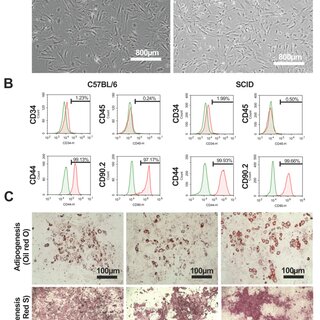
Figure 1. Phenotypes and differentiation potential of C57BL/6 mAdMSCs and SCID mAdMSCs. Morphologies of C57BL/6 mAdMSCs (A, left panel) and SCID mAdMSCs (A, right panel). Flow cytometric analysis of the expression of cell surface markers of C57BL/6 mAdMSCs (B, left panels) and SCID mAdMSCs (B, right panel). Representative images of adipocyte (C, upper left panel) and osteocyte (C, lower left panel) phenotypes of C57BL/6 mAdMSCs cultured in differentiation media. Representative images of adipocyte (C, upper middle panel) and osteocyte (C, lower middle panel)
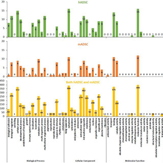
Figure 3. GO analysis of proteins expressed by C57BL/6 mAdMSCs and SCID mAdMSCs. The left, center, and right panels indicate the number of proteins expressed by cultured C57BL/6 and SCID mAdMSCs with emPAI >0, >5, and >10, respectively. The proteins classified in Supplementary Tables 3,5-8 are listed according to the GO categories as follows: growth, immune system process, biological
Figure 4. Functions and features of AdMSCs collected from C57BL/6 and SCID mice associated with different culture conditions: The expression of cell surface markers by mAdMSCs (CD44, CD90.2) was evaluated using immunofluorescence with the same antibodies employed for the flow cytometry experiments (Figure 1B) (A). The proliferation rates of C57BL/6 mAdMSCs and SCID mAdMSCs were not significantly different on days 3 and 5. The data are expressed as the mean ± SD. The y-axis shows the DNA concentration (µg/µL). Day 1: (C57BL/6 mAdMSCs: 5.08 ± 0.91, SCID mAdMSCs: 2.32 ± 0.46, n = 4). Day 3: (C57BL/6 mAdMSCs: 6.92 ± 0.67, SCID mAdMSCs: 5.74 ± 1.79, n = 4). Day 5: (C57BL/6 mAdMSCs: 12.00 ± 4.13, SCID mAdMSCs: 13.34 ± 1.43, n = 4) (B). Real-time PCR analyses (ΔΔCt method) of genes encoding cell surface markers expressed by male SCID mAdMSCs (black bar) and female SCID mAdMSCs (white bar). The expression of the target gene was corrected according to that of HKGs (n = 4) (C). Real-time PCR analysis (ΔΔCt method) of the expression of genes encoding growth factors and antioxidant factors by C57BL/6 mAdMSCs (D, upper panel) and SCID mAdMSCs (D, lower panel) cultured with (black bar) or without (white bar) a gelatin coating (n = 4 each). Realtime PCR analysis (ΔΔCt method) of the expression of genes encoding growth factors and antioxidant factors expressed by C57BL/6 mAdMSCs cultured in the presence (black bar) or absence (white bar) of LPS (E, upper and lower panels, respectively). The levels of the target genes were corrected according to those of HKGs (n = 4) (E, upper panel).
Figure 5. Correlation analysis: A scatter plot showing the correlation (R2 = 0.7475; gray band indicates "R2 = 1") between the normalized values of proteins expressed by C57BL/6 mAdMSCs and SCID mAdMSCs (n = 622). The parallel lines indicate the 95% confidence interval.
Identification of Proteins Differentially Expressed by Adipose-derived Mesenchymal Stem Cells Isolated from Immunodeficient Mice

May 2019

·

107 Reads

·

7 Citations

International Journal of Molecular Sciences

Although cell therapy using adipose-derived mesenchymal stem cells (AdMSCs) regulates immunity, the degree to which cell quality and function are affected by differences in immunodeficiency of donors is unknown. We used liquid chromatography tandem-mass spectrometry (LC MS/MS) to identify the proteins expressed by mouse AdMSCs (mAsMSCs) isolated from normal (C57BL/6) mice and mice with severe combined immunodeficiency (SCID). The protein expression profiles of each strain were 98%-100% identical, indicating that the expression levels of major proteins potentially associated with the therapeutic effects of mAdMSCs were highly similar. Further, comparable levels of cell surface markers (CD44, CD90.2) were detected using flow cytometry or LC MS/MS. and MYO1E, which are associated with immunity, were expressed at higher levels by the SCID mAdMSCs compared with the C57BL/6 mAdMSCs. In contrast, ANXA9, PCBP2, LGALS3, PPP1R14B, and PSMA6, which are also associated with immunity, were more highly expressed by C57BL/6 mAdMSCs than SCID mAdMSCs. These findings implicate these two sets of proteins in the pathogenesis and maintenance of immunodeficiency.


A Liquid Chromatography with Tandem Mass Spectrometry-Based Proteomic Analysis of Primary Cultured Cells and Subcultured Cells Using Mouse Adipose-Derived Mesenchymal Stem Cells

January 2019

·

77 Reads

·

2 Citations

Stem Cells International

Adipose-derived mesenchymal stem cells (MSC-ATs) are representative cell sources for cell therapy. However, how cell stress resulting from passage influences the MSC-AT protein expression has been unclear. In this study, a protein expression analysis was performed by liquid chromatography with tandem mass spectrometry (LC-MS/MS) using mouse primary cultured cells (P0) and cells passaged three times (P3) as samples. A total of 256 proteins were classified as cellular process-related proteins, while 179 were classified as metabolic process-related proteins in P0. These were considered to be adaptive responses of the cells to an in vitro environment. However, seven proteins of growth were identified (Csf1, App, Adam15, Alcam, Tbl1xr1, Ninj1, and Sbds) in P0. In addition, four proteins of antioxidant activity were also identified (Srxn1, Txndc17, Fam213b, and Apoe) in P0. We identified 1139 proteins expressed in both P0 and P3 cells that had their expression decreased to 69.4% in P3 cells compared with P0 cells, but 1139 proteins are very likely proteins that are derived from MSC-AT. The function of MSC-ATs was maintained after three passages. However, the LC-MS/MS analysis data showed that the protein expression was degraded after three passages. MSC-ATs retained about 70% of their protein expression ability in P3 cells.


A Novel Preservation Solution Containing a JNK Inhibitory Peptide Efficiently Improves Islet Yield for Porcine Islet Isolation

December 2018

·

21 Reads

·

13 Citations

Transplantation

Background: For islet transplantation, pancreas preservation in UW solution is associated with disadvantages such as collagenase inhibition, resulting in poor islet yield and islets with poor viability. In this study, we evaluated a novel preservation solution, the extracellular-type c-Jun N-terminal kinase (JNK) inhibitor-containing (EJ) solution. Methods: EJ solution has high sodium-low potassium composition with low viscosity compared to UW solution. Moreover, EJ solution contains a recently developed JNK inhibitor from our laboratory. Results: We first compared the performance of EJ solution with that of UW solution. Islet yield before and after purification was significantly higher in the EJ group than in the UW group. Second, we compared the performance of EJ solution with that of EJ solution without the JNK inhibitor (EJ-J solution). After pancreas preservation in EJ solution, JNK activity was maintained at a relatively low level during islet isolation. Islet yield before and after purification was significantly higher in the EJ group than in the EJ-J group. After islet transplantation into streptozotocin-induced diabetic mice, blood glucose levels reached the normoglycemic range in 61.5% and 7.7% of diabetic mice in the EJ and EJ-J groups, respectively. Moreover, EJ solution exhibited reduced inhibition of collagenase digestion compared to UW solution. Conclusion: Advantages of EJ solution over UW solution were inhibition of JNK activity and reduced collagenase inhibition. EJ solution may therefore be more suitable for islet isolation than UW solution.


Cytokines in adipose-derived mesenchymal stem cells promote the healing of liver disease

November 2018

·

178 Reads

·

23 Citations

World Journal of Stem Cells

Adipose-derived mesenchymal stem cells (ADSCs) are a treatment cell source for patients with chronic liver injury. ADSCs are characterized by being harvested from the patient's own subcutaneous adipose tissue, a high cell yield (i.e., reduced immune rejection response), accumulation at a disease nidus, suppression of excessive immune response, production of various growth factors and cytokines, angiogenic effects, antiapoptotic effects, and control of immune cells via cellcell interaction. We previously showed that conditioned medium of ADSCs promoted hepatocyte proliferation and improved the liver function in a mouse model of acute liver failure. Furthermore, as found by many other groups, the administration of ADSCs improved liver tissue fibrosis in a mouse model of liver cirrhosis. A comprehensive protein expression analysis by liquid chromatography with tandem mass spectrometry showed that the various cytokines and chemokines produced by ADSCs promote the healing of liver disease. In this review, we examine the ability of expressed protein components of ADSCs to promote healing in cell therapy for liver disease. Previous studies demonstrated that ADSCs are a treatment cell source for patients with chronic liver injury. This review describes the various cytokines and chemokines produced by ADSCs that promote the healing of liver disease.


Figure 2. Flow cytometric analysis. hiTS-M cells (passage 14 + 7) and hADSCs (passage 7) were analyzed: (A) hADSCs, CD29; (B) hiTS-M cells, CD29; (C) hADSCs, CD90; (D) hiTS-M cells, CD90; (E) hADSCs, CD44; (F) hiTS-M cells, CD44; (G) hADSCs, CD45; (H) hiTS-M cells, CD45; (I) hADSCs, CD34; and (J) hiTS-M cells, CD34.
Figure 3. Cont.
Induction of Expandable Adipose-Derived Mesenchymal Stem Cells from Aged Mesenchymal Stem Cells by a Synthetic Self-Replicating RNA

November 2018

·

113 Reads

·

5 Citations

International Journal of Molecular Sciences

Adipose-derived mesenchymal stem cells (ADSCs) have attracted attention due to their potential for use in the treatment of various diseases. However, the self-renewal capacity of ADSCs is restricted and their function diminishes during passage. We previously generated induced tissue-specific stem cells from mouse pancreatic cells using a single synthetic self-replicating Venezuelan Equine Encephalitis (VEE)-reprogramming factor (RF) RNA replicon (SR-RNA) expressing the reprogramming factors POU class 5 homeobox 1 (OCT4), Krueppel-like factor 4 (KLF4), Sex determining region Y-box 2 (SOX2), and Glis Family Zinc Finger 1 (GLIS1). This vector was used to generate induced pluripotent stem (iPS) cells. Here, we applied this SR-RNA vector to generate human iTS cells from aged mesenchymal stem cells (hiTS-M cells) deficient in self-renewal that were derived from adipose tissue. These hiTS-M cells transfected with the SR-RNA vector survived for 15 passages. The hiTS-M cells expressed cell surface markers similar to those of human adipose-derived mesenchymal stem cells (hADSCs) and differentiated into fat cells and osteoblasts. Global gene expression profiling showed that hiTS-M cells were transcriptionally similar to hADSCs. These data suggest that the generation of iTS cells has important implications for the clinical application of autologous stem cell transplantation.


Table 1 . Identification of endogenous proteins contained in hADSC; mADSC.
Figure 3. The biological processes, cellular components, and molecular functions of the hADSC, mADSC, and both hADSC and mADSC groups (determined by the Gene Ontology, GO, analysis). The principal component analysis (PCA) of proteome dynamics based on the protein information generated by high-resolution mass spectrometry. The abscissa indicates the biological function, cellular component, and molecular function of the protein. The ordinate indicates the number of proteins identified in the hADSC (top panel), mADSC (middle panel) and both hADSC and mADSC groups (bottom panel). The names of the proteins classified in Table 1 and Supplementary Table S1 are listed according to their detailed biological process (biological adhesion, locomotion, and rhythmic process), cellular component (plasma membrane), and molecular function (antioxidant activity and molecular transducer activity).
A Comparison of Proteins Expressed between Human and Mouse Adipose-Derived Mesenchymal Stem Cells by a Proteome Analysis through Liquid Chromatography with Tandem Mass Spectrometry

November 2018

·

73 Reads

·

11 Citations

International Journal of Molecular Sciences

Adipose-derived mesenchymal stem cells (ADSCs) have become a common cell source for cell transplantation therapy. Clinical studies have used ADSCs to develop treatments for tissue fibrosis, such as liver cirrhosis and pulmonary fibroma. The need to examine and compare basic research data using clinical research data derived from mice and humans is expected to increase in the future. Here, to better characterize the cells, the protein components expressed by human ADSCs used for treatment, and mouse ADSCs used for research, were comprehensively analyzed by liquid chromatography with tandem mass spectrometry. We found that 92% (401 type proteins) of the proteins expressed by ADSCs in humans and mice were consistent. When classified by the protein functions in a gene ontology analysis, the items that differed by >5% between human and mouse ADSCs were “biological adhesion, locomotion” in biological processes, “plasma membrane” in cellular components, and “antioxidant activity, molecular transducer activity” in molecular functions. Most of the listed proteins were sensitive to cell isolation processes. These results show that the proteins expressed by human and murine ADSCs showed a high degree of correlation.


A Liquid Chromatography with Tandem Mass Spectrometry-Based Proteomic Analysis of the Proteins Secreted by Human Adipose-Derived Mesenchymal Stem Cells

October 2018

·

497 Reads

·

20 Citations

Liquid chromatography using a tandem mass spectrometer (LC-MS/MS) is a method of proteomic analysis. A shotgun analysis by LC-MS/MS comprehensively identifies proteins from tissues and cells with high resolution. The hepatic function of mice with acute hepatitis following the intraperitoneal administration of CCL4 was improved by the tail vein administration of the culture conditional medium (CM) of human mesenchymal stem cells from adipose tissue (hMSC-AT). In this study, a secreted protein expression analysis of hMSC-AT was performed using LC-MS/MS; 128 proteins were identified. LC-MS/MS showed that 106 new functional proteins and 22 proteins (FINC, PAI1, POSTN, PGS2, TIMP1, AMPN, CFAH, VIME, PEDF, SPRC, LEG1, ITGBL, ENOA, CSPG2, CLUS, IBP4, IBP7, PGS1, IBP2, STC2, CTHR1, CD9) were previously reported in hMSC-AT-CMs. In addition, various proteins associated with growth (SAP, SEM7A, PTK7); immune system processes (CO1A2, CO1A1, CATB, TSP1, GAS6, PTX3, C1 S, SEM7A, G3P, PXDN, SRCRL, CD248, SPON2, ENPP2, CD109, CFAB, CATL1, MFAP5, MIF, CXCL5, ADAM9, CATK); and reproduction (MMP2, CATB, FBLN1, SAP, MFGM, GDN, CYTC) were identified in hMSC-AT-CMs. These results indicate that a comprehensive expression analysis of proteins by LC-MS/MS is useful for investigating new factors associated with cellular components, biological processes, and molecular functions.


Citations (85)


... Currently, some scholars revealed ANXA3 was involved in immune and in ammatory progress [11,12]. ...

Reference:

Evaluation and validation of the increased annexin A3 (ANXA3) as a novel biomarker to predict sepsis in critically ill patients
Identification of Proteins Differentially Expressed by Adipose-derived Mesenchymal Stem Cells Isolated from Immunodeficient Mice

International Journal of Molecular Sciences

... The iTS cells derived from pancreatic tissue (iTS-P cells), which express several endoderm/pancreatic markers, differentiate into insulin-expressing cells more efficiently than ES/iPS cells. More important, iTS cells do not form teratomas when subcutaneously transplanted into immunodeficient mice [5][6][7][8] . Therefore, iTS cells provide important advantages over ES/iPS cells. ...

Induction of expandable tissue-specific progenitor cells from human pancreatic tissue through transient expression of reprogramming factors

Molecular Therapy — Methods & Clinical Development

... According to the results of recent studies, injecting MSC-conditioned medium (CM) into the anterior cruciate ligament transection rat model is sufficient to mediate cartilage regeneration function [11,[15][16][17]; thus, interest in efficacious knee OA treatments using MSC-derived paracrine factors has increased [14,[18][19][20][21]. However, although several studies have actively investigated paracrine factors secreted by each type of MSC obtained from various tissues under general culture conditions [22][23][24], it is still unclear which paracrine factors affect the regeneration of damaged cartilage. ...

A Liquid Chromatography with Tandem Mass Spectrometry-Based Proteomic Analysis of Primary Cultured Cells and Subcultured Cells Using Mouse Adipose-Derived Mesenchymal Stem Cells

... On the other hand, the number of islets decrease after islet purification. This is due to the presence of islets in less pure fractions after islet purification and/or the death of islets by various stressors such as exposure to cytokines/chemokines and mechanical stress [12][13][14][15]. ...

A Novel Preservation Solution Containing a JNK Inhibitory Peptide Efficiently Improves Islet Yield for Porcine Islet Isolation
  • Citing Article
  • December 2018

Transplantation

... There are still some other common SCs, but they may be less common in neonatal disease research applications; examples include the following: (1) In vitro and in vivo studies have shown that human adipose-derived MSCs (hADSCs) are pluripotent and can differentiate into mesoderm-derived cells, including fat cells, chondrocytes, and osteoblasts [34]. hADSCs enter the surrounding environment through secretion or production of a range of cytokines, growth factors, nucleic acids (miRNAs), and other macromolecules through microvesicles, altering histobiology, stimulating tissue-resident SCs, altering immune cell activity, and mediating treatment outcomes [35]. Their advantages include abundant sources, easy tissue collection and cell isolation, and therapeutic potential [34]. ...

Cytokines in adipose-derived mesenchymal stem cells promote the healing of liver disease

World Journal of Stem Cells

... The comprehensive expression analysis of secreted proteins in supernatants of Rab37-ADSCs using LC-MS/MS was performed according to a previously reported method [33,34]. In this study, the data obtained by LC-MS/MS were quantified according the theoretical value (emPAI), estimated based on the function of the Scaffold software program [33,43]. ...

A Comparison of Proteins Expressed between Human and Mouse Adipose-Derived Mesenchymal Stem Cells by a Proteome Analysis through Liquid Chromatography with Tandem Mass Spectrometry

International Journal of Molecular Sciences

... The aging-related cellular phenotypes such as mitochondria function and membrane integrity could be reversed through de-differentiation into a pluripotent state, often through transcriptive reprogramming via overexpressing the key stemness markers including octamer-binding transcription factor 4 (Oct4), the sex-determining region Y-box 2 (Sox2) and kruppel-like factor 4 (Klf4) [171,172]. Noguchi et al. reported that the transfection of Venezuelan Equine Encephalitis (Vee)-reprogramming factor (Rf) RNA replicon (SR-RNA) co-expressing Oct4, Sox2 and Klf4 can be used to generate the reprogramed iPSCs with high proliferation ability [173]. However, while these iPSCs reprogrammed from MSCs resemble the profiles of somatic/embryonic stages, this reestablishment of the self-renewal and pluripotent stage is not a viable therapeutic strategy in vivo, as the direct injection of pluripotent cells leads to cancer and teratoma formation in mice [174,175]. ...

Induction of Expandable Adipose-Derived Mesenchymal Stem Cells from Aged Mesenchymal Stem Cells by a Synthetic Self-Replicating RNA

International Journal of Molecular Sciences

... However, identifying these cells can be a challenging task, as even advanced techniques such as single-cell RNA sequencing have not been able to fully characterize the distinct cell clusters within the ovarian stroma (Fan et al., 2019). Alternatively, the identification of the bioactive factors in the spent media could be performed by enzyme-linked immunosorbent assay or liquid chromatography with tandem mass spectrometry (Chiswick et al., 2012;Nakashima et al., 2018) to reveal the interplay between OSCs and follicles. ...

A Liquid Chromatography with Tandem Mass Spectrometry-Based Proteomic Analysis of the Proteins Secreted by Human Adipose-Derived Mesenchymal Stem Cells

... Adipose tissue was harvested from the inguinal region of NOD mice under general anesthesia in a sterile environment. The isolation of ADSCs was performed as previously described 52 . Briefly, the adipose tissue was stored in Hank's Balanced Salt Solution (HBSS) and vigorously washed with HBSS. ...

A Comparison of the Preservation of Mouse Adipose Tissue-Derived Mesenchymal Stem Cells Using the University of Wisconsin Solution and Hank's Balanced Salt Solution

... The iTS cells derived from pancreatic tissue (iTS-P cells), which express several endoderm/pancreatic markers, differentiate into insulin-expressing cells more efficiently than ES/iPS cells. More important, iTS cells do not form teratomas when subcutaneously transplanted into immunodeficient mice [5][6][7][8] . Therefore, iTS cells provide important advantages over ES/iPS cells. ...

Characterization of induced tissue-specific stem cells from pancreas by a synthetic self-replicative RNA

Scientific Reports