David Mazaud's research while affiliated with Collège de France and other places

What is this page?


This page lists the scientific contributions of an author, who either does not have a ResearchGate profile, or has not yet added these contributions to their profile.

It was automatically created by ResearchGate to create a record of this author's body of work. We create such pages to advance our goal of creating and maintaining the most comprehensive scientific repository possible. In doing so, we process publicly available (personal) data relating to the author as a member of the scientific community.

If you're a ResearchGate member, you can follow this page to keep up with this author's work.

If you are this author, and you don't want us to display this page anymore, please let us know.

Publications (36)


Epg5 links proteotoxic stress due to defective autophagic clearance and epileptogenesis in Drosophila and Vici Syndrome patients
  • Preprint
  • File available

April 2024

·

50 Reads

·

·

Simon Lowe

·

[...]

·

Epilepsy is a common neurological condition that arises from dysfunctional neuronal circuit control due to either acquired or innate disorders. Autophagy is an essential neuronal housekeeping mechanism, which causes severe proteotoxic stress when impaired. Autophagy impairment has been associated to epileptogenesis through a variety of molecular mechanisms. Vici Syndrome (VS) is the paradigmatic congenital autophagy disorder in humans due to recessive variants in the ectopic P-granules autophagy tethering factor 5 (EPG5) gene that is crucial for autophagosome-lysosome fusion and ultimately for effective autophagic clearance. VS is characterized by a wide range of neurodevelopmental, neurodegenerative, and neurological features, including epilepsy. Here, we used Drosophila melanogaster to study the importance of epg5 in development, ageing, and seizures. Our data indicate that proteotoxic stress due to impaired autophagic clearance and seizure-like behaviors correlate and are commonly regulated, suggesting that seizures occur as a direct consequence of proteotoxic stress and age-dependent neurodegenerative progression in epg5 Drosophila mutants, in the absence of evident neurodevelopmental abnormalities. We provide complementary evidence from EPG5-mutated patients demonstrating an epilepsy phenotype consistent with Drosophila predictions and propose autophagy stimulating diets as a feasible approach to control EPG5-related pharmacoresistant seizures.

Download
Share

Figure 1. High astrocyte density in the visual layer of the superior colliculus. (a) Coronal sections of the visual layer (VL) of the superior colliculus (SC, left panel), primary visual cortex (V1, middle panel) and hippocampal CA1 (CA1, right panel). Regions were identified with a myelin basic protein staining (MBP, top panels). For the hippocampus, the stratum oriens (s.o.), stratum pyrimidale (s.p.) and stratum radiatum (s.r.) are indicated. Representative images for neuron (middle panels) and astrocyte densities (bottom panels). (b, c) Quantification of neuron (b) and astrocyte (c) density in the SC (n=12), V1 (n=9) and CA1 (n=10). One-way ANOVA (neuron: F(2,28) = 87.75), p < 0.0001, with Tukey's multiple comparisons ***p < 0.0001); astrocyte: F(2,28) = 67.74, p<0.0001, with Tukey's multiple comparisons *p < 0.0001, ***p < 0.0001). (d) Quantification of astrocyte to neuron density ratio. One-way ANOVA (F(2,28) = 64.15, p < 0.0001, with Tukey's multiple comparisons ***p < 0.0001). n represents the number of brain coronal slices that were analyzed from 3 mice. Scale bar, 25µm. n=3 mice; data are shown as mean ± SEM.
Figure 3. Low intracellular diffusion in astrocytes from the visual layer of the superior colliculus. (a) Schematic overview showing the approach used to quantify intracellular diffusion in astrocytes from different brain areas, the visual layer of the superior colliculus (SC), the primary visual cortex (V1) and the CA1 region of the hippocampus (CA1). Cytoplasmic eGFP expressed by astrocytes was bleached by high laser power line scanning. Recovery occurred when unbleached GFP entered the scanned area. Fluorescence Recovery After Photobleaching (FRAP) was calculated by dividing the bleaching factor (ΔFb) by the recovery fraction (ΔFr). (b) FRAP was significantly reduced in the SC (n =8) compared to V1 (n = 11) and CA1 (n = 11) (One-way ANOVA, F(2,27) = 9.806, p = 0.0006, with Tukey's multiple comparisons *p < 0.05, ***p < 0.001). (c) Diffusion speed in the soma was similar (One-way ANOVA, F(2,26) = 0.4528, p = 0.6407). (d) The difference in intracellular diffusion speed arises from the processes, as FRAP is lower in the SC (One-way ANOVA, F(2,27) = 9.840, p = 0.0006, with Tukey's multiple comparisons *p < 0.05, ***p < 0.0001). Scale bar, 10 µm. n represents the number of cells analysed from 3 mice. Data are shown as mean ± SEM.
Figure 4. Tight astroglial coverage of synapses in the visual layer of the superior colliculus. (a) Schematic illustrating the method used to measure the distances between astrocytic processes and synapses (left, top panel) with super-resolution STED microscopy combining an excitation and a depletion laser (left, bottom panel). Using this method, synaptic boutons can be accurately distinguished (right, lower panel), as shown in the representative images compared to confocal microscopy (right, top panel). Scale bar, 0.1µm. (b) Top: representative confocal images of astrocytes labelled with eGFP in the visual layer of the superior colliculus (SC), primary visual cortex (V1) and hippocampal CA1 (CA1), together with VGLUT1 and Homer to visualize pre-and postsynaptic elements, respectively. Scale bar, 10µm. Bottom: representative STED images of an astrocytic process with a synapse in each brain structure. Scale bar, 0.1 µm. (c) Cumulative frequency of the distances between astrocyte process and synapse. Kolmogorov-Smirnov (SC (n = 6710 synapses) vs V1 (n = 8702 synapses): ***p < 0.0001; SC vs CA1 (n = 9837 synapses): ***p < 0.0001; V1 vs CA1: ***p < 0.0001). (d) Frequency distributions of distance between processes and synapses. Short distances were defined as distances up to 210 nm. (e) Quantification of the number of short distance processes (SC: n = 16, V1: n = 17, CA1: n = 13, One-way ANOVA, F(2,43) = 28.45, p < 0.0001, with Tukey's multiple comparisons ***p < 0.0001). n is represented as number of astrocytes, which are acquired from 4 mice, unless differently indicated, data are shown as mean ± SEM.
Astrocytes in the retinorecipient superior colliculus display unique cellular and structural properties

August 2022

·

155 Reads

Astrocytes have long been considered to be a largely homogeneous cell population. Recent studies however suggest that astrocytes are highly adapted to the local neuronal circuitry. Glucose utilization in the retinorecipient superior colliculus (SC) is one of the highest in the brain. Since metabolic support to neurons is a major function of astrocytes, they could be of particular relevance in this region and display specific features. However, little is known about astrocytes and their interactions with neurons in this multisensory brain area. We thus here investigated region-specific cellular and structural properties of astrocytes in the visual layer of the SC. Using morphological reconstructions, fluorescent recovery after photobleaching (FRAP) and superresolution imaging, we found that astrocytes from the visual layers of the SC are highly distinct with a higher cellular density, a more complex morphology and a stronger proximity to synapses compared to astrocytes from the primary visual cortex and the hippocampus. These data point to astroglial diversity and specialization within neural circuits integrating sensory information in the adult brain.


Correction: Fast calcium transients in dendritic spines driven by extreme statistics

May 2022

·

36 Reads

PLOS BiologyPLOS Biology

[This corrects the article DOI: 10.1371/journal.pbio.2006202.].


Nanoscale molecular architecture controls calcium diffusion and ER replenishment in dendritic spines

September 2021

·

107 Reads

·

14 Citations

Science Advances

Dendritic spines are critical components of neuronal synapses as they receive and transform synaptic inputs into a succession of calcium-regulated biochemical events. The spine apparatus (SA), an extension of smooth endoplasmic reticulum, regulates slow and fast calcium dynamics in spines. Calcium release events deplete SA calcium ion reservoir rapidly, yet the next cycle of signaling requires its replenishment. How spines achieve this replenishment without triggering calcium release remains unclear. Using computational modeling, calcium and STED superresolution imaging, we show that the SA replenishment involves the store-operated calcium entry pathway during spontaneous calcium transients. We identified two main conditions for SA replenishment without depletion: a small amplitude and a slow timescale for calcium influx, and a close proximity between SA and plasma membranes. Thereby, spine’s nanoscale organization separates SA replenishment from depletion. We further conclude that spine’s receptor organization also determines the calcium dynamics during the induction of long-term synaptic changes.


The many ways astroglial connexins regulate neurotransmission and behavior

June 2021

·

75 Reads

·

15 Citations

Glia

Astrocytes have emerged as major players in the brain, contributing to many functions such as energy supply, neurotransmission, and behavior. They accomplish these functions in part via their capacity to form widespread intercellular networks and to release neuroactive factors, which can modulate neurotransmission at different levels, from individual synapses to neuronal networks. The extensive network communication of astrocytes is primarily mediated by gap junction channels composed of two connexins, Cx30 and Cx43, which present distinct temporal and spatial expression patterns. Yet, astroglial connexins are also involved in direct exchange with the extracellular space via hemichannels, as well as in adhesion and signaling processes via unconventional nonchannel functions. Accumulating evidence indicate that astrocytes modulate neurotransmission and behavior through these diverse connexin functions. We here review the many ways astroglial connexins regulate neuronal activity from the molecular level to behavior.


Nanoscale organization and calcium influx in dendritic spines guarantee ER store refilling without depletion

June 2021

·

32 Reads

Dendritic spines are critical components of the neuronal synapse as they receive and transform the synaptic input into a succession of biochemical events regulated by calcium signaling. The spine apparatus (SA), an extension of smooth endoplasmic reticulum (ER), regulates slow and fast calcium dynamics in spines. Calcium release events from SA result in a rapid depletion of calcium ion reservoir, yet the next cycle of signaling requires replenishment of SA calcium stores. How dendritic spines achieve this without triggering calcium release remains unclear. Using computational modeling, calcium and STED super-resolution imaging, we showed that the refilling of calcium-deprived SA involves store-operated calcium entry during spontaneous calcium transients in spine heads. We identified two main conditions that guarantee SA replenishment without depletion: (1) a small amplitude and slow timescale for calcium influx, and (2) a close proximity between SA and plasma membranes. Thereby, molecular nano-organization creates the conditions for a clear separation between SA replenishment and depletion. We further conclude that the nanoscale organization of SA receptors underlies the specificity of calcium dynamics patterns during the induction of long-term synaptic changes.


Figure 1. The Perisynaptic Processes of Hippocampal Astrocytes Synthesize Proteins and Contain Protein Maturation Organelles (A) Representative confocal microscopy image of the FISH detection of Slc1a2 mRNAs (red dots) in a dorsal hippocampal CA1 astrocyte immunolabeled for GFAP (green). Nuclei were stained with DAPI (blue). (B) AstroDot analysis of the distribution of Slc1a2 mRNA in somata, large (>0.3 mm) and fine (<0.3 mm) GFAP-immunolabeled processes, and GFAP-negative processes of CA1 astrocytes (n = 55 cells in three mice). (C) Detection of CA1 astrocyte ribosomes and local translation events in PAPs by puromycylation (+PMY, with puromycin) in Aldh1l1:L10a-eGFP transgenic mice. Astrocyte ribosomes were immunolabeled for GFP (green) and PMY (red). Synapses were co-immunolabeled for VGluT1 (pre-synapse) (gray) and Homer1 (postsynapse) (blue). The white square is a magnified view (103) of an astrocyte translation event near synapses. The astrocyte domain is indicated by a dotted line. (D) Quantification of the percentage of Homer1/VGluT1-positive synapses within 1 mm of a L10A-eGFP signal. n = 128 cells in three mice. (legend continued on next page) Cell Reports 32, 108076, August 25, 2020 3
Figure 4. Comparison of Enriched Ribosome-Bound mRNA Levels in PAPs versus PvAPs (A) Flowchart for the comparison of a selection of enriched mRNAs in PAPs versus PvAPs. Ribosome-bound mRNAs were isolated using TRAP from Aldh1l1:L10a-eGFP whole hippocampal synaptogliosomes or purified gliovascular units. The raw qPCR data are given in Table S3. (B) qPCR results for Fth1, Rplp1, and Ezr, demonstrating their enrichment in PAPs versus PvAPs. *p % 0.05, one-tailed Mann-Whitney test. The data are expressed as mean ± SEM (n = 3).
Figure 6. Local Translation Changes in PAPs after Fear Conditioning (A) Flowchart for the fear-conditioning protocol, followed by analyses of mRNAs and proteins in astrocytes and PAPs from the dorsal hippocampus. (B) AstroDot analysis of the density of PAP-enriched mRNAs encoding RACK1 (Gnb2l1), FTL1, FTH1, eEF1A1 MDM2, and CCND2 in GFAP-immunolabeled astrocytic processes in the dorsal hippocampus of control mice and fear-conditioned mice. *p < 0.05, ***p < 0.001, ****p < 0.0001, and ns (not significant) in an unpaired two-tailed t test; n R 35 cells from three mice per condition. The data are presented as mean ± SEM, and the raw data are given in Table S4.
Local Translation in Perisynaptic Astrocytic Processes Is Specific and Changes after Fear Conditioning

August 2020

·

346 Reads

·

52 Citations

Cell Reports

Local translation is a conserved mechanism conferring cells the ability to quickly respond to local stimuli. In the brain, it has been recently reported in astrocytes, whose fine processes contact blood vessels and synapses. Yet the specificity and regulation of astrocyte local translation remain unknown. We study hippocampal perisynaptic astrocytic processes (PAPs) and show that they contain the machinery for translation. Using a refined immunoprecipitation technique, we characterize the entire pool of ribosome-bound mRNAs in PAPs and compare it with the one expressed in the whole astrocyte. We find that a specific pool of mRNAs is highly polarized at the synaptic interface. These transcripts encode an unexpected molecular repertoire, composed of proteins involved in iron homeostasis, translation, cell cycle, and cytoskeleton. Remarkably, we observe alterations in global RNA distribution and ribosome-bound status of some PAP-enriched transcripts after fear conditioning, indicating the role of astrocytic local translation in memory and learning.


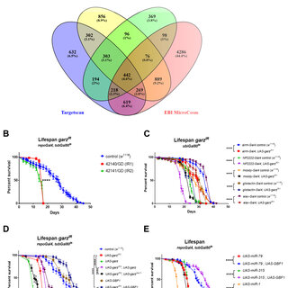
Figure 1. Effects of garz knock-down in adult glial cell. (A) Venn diagram referring to the data in Table 2 and illustrating the overlap between the four different databases used to predict gene targets of the miRNAs whose expression in the adult glia resulted in a significant reduction in fly lifespan. Only 520 target genes are in common among all four databases, garz falls in this group. A remarkably large number of genes as targets were uniquely predicted by the MicroCosm database. (B) Two RNAi lines against garz bring about a very significant reduction in fly lifespan in comparison to controls, when expressed in all adult glia. N=60 for each genotype, Error bars SEM, pairwise comparisons: Log-rank (Mantel-Cox) test. The full dataset can be accessed at DOI 10.17605/OSF.IO/8E3NS as part of Table 3. (C) Knock-down of garz in sub-populations of glial cells, astrocyte-like (alrm-Gal4), Cortex glia (NP2222-Gal4), sub-perineural glia (moody-Gal4), perineural and PNS glia (gliotactin-gal4) or in neurons (elav-Gal4) brings about a significant reduction in lifespan in comparison to controls. N=60 for each genotype, Error bars SEM, pairwise comparisons: Log-rank (Mantel-Cox) test. The full dataset can be accessed at DOI 10.17605/OSF. IO/HQCDG. (D) Lifespan reduction due to RNAi against garz in adult glia is rescued by an exogenous UAS-garz transgene and by a transgene expressing the human orthologue GBF1 under UAS control. Note that overexpression of garz in an otherwise wt background is highly detrimental to fly lifespan, whereas overexpression of GBF1 in a wt background has no adverse effects. Mutations leading to a non-functional Sec7 domain eliminate or drastically reduce the ability of garz or GBF1 transgenes to rescue fly lifespan. N=60 for each genotype, Error bars SEM, pairwise comparisons: Log-rank (Mantel-Cox) test. The full dataset can be accessed at DOI 10.17605/OSF.IO/ 5RGEF. (F) Co-expression of human GBF1 significantly extends the short lifespan caused by overexpression of miR-1, miR-79 and miR-315 in adult glia. N=60 for each genotype, Error bars SEM, pairwise comparisons: Log-rank (Mantel-Cox) test. The full dataset can be accessed at DOI 10.17605/OSF.IO/B37DF.
Figure 2. Knock down of garz in adult glial cells leads to significant impairment of fly motor functions. All data in this figure represent a grouping of two independent experiments with a total number of flies analysed (N) of 35-40. Error bars represent SEM in all graphs. Untreated track data can be accessed at DOI 10.17605/OSF.IO/UNJX7. (A) Stimulus response curve for control flies (black), garz RNAi (red) and co-expression of GBF1 and garz RNAi (green). The graph is an average of 6 tracks for each of the stimuli received at 15 min intervals (See Methods). All genotypes also include repo-Gal4 and ubi-Gal80 ts to express the transgenes in all adult glia. In control flies the presence of tub-Gal80 blocks any expression of UAS-transgenes. The graph to the right reports the mean amplitude of the response to a train of stimuli, which is significantly reduced by RNAi against garz, and this reduction is reverted to normal level by co-expression of human GBF1. One-way ANOVA, Dunnett's multiple comparisons post hoc test. (B) Average speed analysis of the same flies as in A. RNAi against garz significantly slows down fly motility and this is rescued by human GBF1. One-way ANOVA, Dunnett's multiple comparisons post hoc test. (C) Mean bout length analysis of the same flies as in A. No significant difference is detected in this parameter. One-way ANOVA, Dunnett's multiple comparisons post hoc test. (D) Mean interbout interval analysis of the same flies as in A. RNAi against garz significantly increases the time spent in inactivity by flies and this is rescued by human GBF1. One-way ANOVA, Dunnett's multiple comparisons post hoc test.
Figure 3. Sub-cellular dysfunctions caused by garz knock-down in adult glial cell. (A) Representative single confocal sections of adult fly brains stained for DAPI (blue), GFP (green), Repo (magenta) and Ref (2)P (red). Pan glial knock-down of garz with repo-Gal4 and ubi-Gal80 ts leads to abnormal distribution of the plasma membrane targeted CD8-GFP protein (expressed from a UAS-CD8-GFP transgene in all glial cells) leading to gaps and blebs (arrows, see also Videao1 and 2), and to accumulation of Ref(2)P puncta (arrowheads). The full dataset can be accessed at DOI 10.17605/OSF.IO/96TS3. (B) Representative single confocal section of adult fly brains stained for DAPI (blue), GFP (green) and Repo (red). The GFP signal also in back and white (lower panels) is due to the presence of a UAS-mitoGFP transgenes and detects mitochondria. (C) Quantification of mitochondria parameters based on the GFP signal in B. Pan glial knock-down of garz with repo-Gal4 and ubi-Gal80 ts leads to significant increases in the volume, surface area and sphericity of mitochondria. Mann-Whitney non-parametric test. N=300 objects, randomly selected from 4 brains. Error bars represent SEM. The full dataset can be accessed at DOI 10.17605/OSF.IO/EXMTG.
A miRNA screen procedure identifies garz as an essential factor in adult glia functions and validates Drosophila as a beneficial 3Rs model to study glial functions and GBF1 biology

July 2020

·

74 Reads

Invertebrate glia performs most of the key functions controlled by mammalian glia in the nervous system and provides an ideal model for genetic studies of glial functions. To study the influence of adult glial cells in ageing we have performed a genetic screen in Drosophila using a collection of transgenic lines providing conditional expression of micro-RNAs (miRNAs). Here, we describe a methodological algorithm to identify and rank genes that are candidate to be targeted by miRNAs that shorten lifespan when expressed in adult glia. We have used four different databases for miRNA target prediction in Drosophila but find little agreement between them, overall. However, top candidate gene analysis shows potential to identify essential genes involved in adult glial functions. One example from our top candidates’ analysis is gartenzwerg ( garz ). We establish that garz is necessary in many glial cell types, that it affects motor behaviour and, at the sub-cellular level, is responsible for defects in cellular membranes, autophagy and mitochondria quality control. We also verify the remarkable conservation of functions between garz and its mammalian orthologue, GBF1, validating the use of Drosophila as an alternative 3Rs-beneficial model to knock-out mice for studying the biology of GBF1, potentially involved in human neurodegenerative diseases.


Figure 1. Effects of garz knock-down in adult glial cell. (A) Venn diagram referring to the data in Table 6 and illustrating the overlap between the four different databases used to predict gene targets of the miRNAs whose expression in the adult glia resulted in a significant reduction in fly lifespan. Only 520 target genes are in common among all four databases, garz falls in this group. A remarkably large number of genes as targets were uniquely predicted by the MicroCosm database. (B) Two RNAi lines against garz bring about a very significant reduction in fly lifespan in comparison to controls, when expressed in all adult glia. N=60 for each genotype, Error bars SEM, pairwise comparisons: Log-rank (Mantel-Cox) test. The full dataset can be accessed at DOI 10.17605/OSF.IO/8E3NS as part of Table 7. (C) Knock-down of garz in sub-populations of glial cells, astrocyte-like (alrm-Gal4), Cortex glia (NP2222-Gal4), sub-perineural glia (moody-Gal4), perineural and PNS glia (gliotactin-gal4) or in neurons (elav-Gal4) brings about a significant reduction in lifespan in comparison to controls. N=60 for each genotype, Error bars SEM, pairwise comparisons: Log-rank (Mantel-Cox) test. The full dataset can be accessed at DOI 10.17605/OSF. IO/HQCDG. (D) Lifespan reduction due to RNAi against garz in adult glia is rescued by an exogenous UAS-garz transgene and by a transgene expressing the human orthologue GBF1 under UAS control. Note that overexpression of garz in an otherwise wt background is highly detrimental to fly lifespan, whereas overexpression of GBF1 in a wt background has no adverse effects. Mutations leading to a non-functional Sec7 domain eliminate or drastically reduce the ability of garz or GBF1 transgenes to rescue fly lifespan. N=60 for each genotype, Error bars SEM, pairwise comparisons: Log-rank (Mantel-Cox) test. The full dataset can be accessed at DOI 10.17605/OSF.IO/ 5RGEF. (F) Co-expression of human GBF1 significantly extends the short lifespan caused by overexpression of miR-1, miR-79 and miR-315 in adult glia. N=60 for each genotype, Error bars SEM, pairwise comparisons: Log-rank (Mantel-Cox) test. The full dataset can be accessed at DOI 10.17605/OSF.IO/B37DF.
Figure 3. Sub-cellular dysfunctions caused by garz knock-down in adult glial cell. (A) Representative single confocal sections of adult fly brains stained for DAPI (blue), GFP (green), Repo (magenta) and Ref (2)P (red). Pan glial knock-down of garz with repo-Gal4 and ubi-Gal80 ts leads to abnormal distribution of the plasma membrane targeted CD8-GFP protein (expressed from a UAS-CD8-GFP transgene in all glial cells) leading to gaps and blebs (arrows, see also Videao1 and 2), and to accumulation of Ref(2)P puncta (arrowheads). The full dataset can be accessed at DOI 10.17605/OSF.IO/96TS3. (B) Representative single confocal section of adult fly brains stained for DAPI (blue), GFP (green) and Repo (red). The GFP signal also in back and white (lower panels) is due to the presence of a UAS-mitoGFP transgenes and detects mitochondria. (C) Quantification of mitochondria parameters based on the GFP signal in B. Pan glial knock-down of garz with repo-Gal4 and ubi-Gal80 ts leads to significant increases in the volume, surface area and sphericity of mitochondria. Mann-Whitney non-parametric test. N=300 objects, randomly selected from 4 brains. Error bars represent SEM. The full dataset can be accessed at DOI 10.17605/OSF.IO/EXMTG.
A miRNA screen procedure identifies garz as an essential factor in adult glia functions and validates Drosophila as a beneficial 3Rs model to study glial functions and GBF1 biology

May 2020

·

85 Reads

Invertebrate glia performs most of the key functions controlled by mammalian glia in the nervous system and provides an ideal model for genetic studies of glial functions. To study the influence of adult glial cells in ageing we have performed a genetic screen in Drosophila using a collection of transgenic lines providing conditional expression of micro-RNAs (miRNAs). Here, we describe a methodological algorithm to identify and rank genes that are candidate to be targeted by miRNAs that shorten lifespan when expressed in adult glia. We have used four different databases for miRNA target prediction in Drosophila but find little agreement between them, overall. However, top candidate gene analysis shows potential to identify essential genes involved in adult glial functions. One example from our top candidates’ analysis is gartenzwerg ( garz ). We establish that garz is necessary in many glial cell types, that it affects motor behaviour and, at the sub-cellular level, is responsible for defects in cellular membranes, autophagy and mitochondria quality control. We also verify the remarkable conservation of functions between garz and its mammalian orthologue, GBF1, validating the use of Drosophila as an alternative 3Rs-beneficial model to knock-out mice for studying the biology of GBF1, potentially involved in human neurodegenerative diseases.


Local translation in perisynaptic astrocytic processes is specific and regulated by fear conditioning

January 2020

·

135 Reads

·

1 Citation

Local translation is a conserved molecular mechanism conferring cells the ability to quickly respond to local stimuli. It not only permits cells with complex morphology to bypass somatic protein synthesis and transport, but also contributes locally to the establishment of molecular and functional polarity. In the brain, local translation has been extensively studied in neurons and has only been recently reported in astrocytes, whose fine processes contact both blood vessels and synapses. Yet the specificity and regulation of astrocyte local translation remain unknown. Here, we studied hippocampal perisynaptic astrocytic processes (PAPs) and show that they contain all the machinery for translation. Using our recently refined polysome immunoprecipitation technique, we then characterized the pool of polysomal mRNAs in PAPs, referred to as the PAPome, and compared it to the one found in the whole astrocyte. We found that the PAPome encoded an unexpected molecular repertoire, mostly composed of cytoplasmic proteins and of proteins involved in iron homeostasis, translation, cell cycle and cytoskeleton. Among them, ezrin (Ezr), ferritin heavy chain 1 (Fth1) and 60S acidic ribosomal protein1 (Rplp1) were enriched in PAPs compared to perivascular astrocytic processes, indicating that local translation differs at these two interfaces. Remarkably, PAPs were also enriched in transcripts coding for proteins involved in learning and memory, such as ferritin (Ftl1 and Fth1), G1/S-specific cyclin-D2 (Ccnd2), E3 ubiquitin-protein ligase (Mdm2) , Receptor of activated protein C kinase 1 (Gnb2l1) and Elongation factor 1-alpha 1 (Eef1a1). To address their regulation in a physiological context, we assessed their local translation after fear conditioning. We found alterations in their density and/or distribution in astrocytes as well as a drop in their translation specifically in PAPs. In all, our results reveal an unexpected molecular repertoire of hippocampal PAPs, which is regulated by local translation during learning and memory processes.


Citations (8)


... 85 In neurons, both STIM and ORAI proteins are detected at synapses and preferentially localize to SA-bearing spines. 55,86 Loss of STIM2 or ORAI1 induces an impairment in dendritic spine maturation [87][88][89][90] and attenuates LTP. 91 In addition, STIM2 may directly interact with GluA1-containing AMPARs to control their plasticity-induced surface expression. ...

Reference:

Cell-surface receptor-mediated regulation of synaptic organelle distribution controls dendritic spine maturation
Nanoscale molecular architecture controls calcium diffusion and ER replenishment in dendritic spines

Science Advances

... For example, astrocytes directly manipulate synaptic network formation and function through various signaling molecules (Saint-Martin and Goda, 2022;Farizatto and Baldwin, 2023), remodel the extracellular matrix (Tewari et al., 2022;Dzyubenko and Hermann, 2023), perform phagocytosis (Park and Chung, 2023), and regulate extracellular ions and neurotransmitters during neural activity (Andersen and Schousboe, 2023;Purushotham and Buskila, 2023). Beside neurons, astrocytes also conduct intercellular communication not only among themselves (Barber et al., 2021;Mazaud et al., 2021), but also to the neurovasculature (Diaz-Castro et al., 2023;Lia et al., 2023), other glia (e.g., oligodendrocytes and microglia; Liu et al., 2023), and peripheral immune cells (Han et al., 2021). In neuropathological conditions such as injury, inflammation, and neurodegenerative diseases, astrocyte signaling cues can become dysregulated and thus may be targets for therapeutic intervention (Brandebura et al., 2023;Lawrence et al., 2023;Patani et al., 2023;Qian et al., 2023). ...

The many ways astroglial connexins regulate neurotransmission and behavior
  • Citing Article
  • June 2021

Glia

... Given that manipulating astrocyte functions bidirectionally modulates drug-seeking behaviors, these cells may play a more fundamental role in the cycle of addiction than previously believed. Additionally, while not discussed in this review, exciting research indicates that post-translational and protein-trafficking mechanisms in astrocytes are also important in regulating complex behaviors Mazare et al. 2020;Farrell et al. 2023). How substances of abuse change the regulation of the astrocyte transcriptome, at the transcriptional and epigenetic levels, remains elusive but could provide important insights to the biological underpinnings of addiction. ...

Local Translation in Perisynaptic Astrocytic Processes Is Specific and Changes after Fear Conditioning

Cell Reports

... These mechanisms also vary according to the cell type and spatial organization. For example, single cell comparative measurements of mRNA level between the soma and dendritic parts of neurons have surprisingly revealed higher abundance of some specific ribosome proteins in the dendritic region [62] (similar observation was also found in glial cells [63]). An interesting hypothesis, suggested by cell imaging, is that the ribosomal proteins in dendrites actually join pre-existing ribosomes, to maintain translation activity in axons [64], far from the nucleus where the ribosome is assembled. ...

Local translation in perisynaptic astrocytic processes is specific and regulated by fear conditioning

... Overall, it can be assumed that the functions of synaptic and extrasynaptic NMDA receptors may differ significantly. While the former are targeted at the influx of Ca 2+ ions and further activation of various pathways of synaptic plasticity [45,46], the latter may be directly related to the regulation of the delivery of synaptic signals to the soma [38,47], which will be discussed below. ...

Fast calcium transients in dendritic spines driven by extreme statistics

... We then found that re-expressing Drosophila Aprt selectively in neurons in Aprt 5 background partially rescued the SING phenotype of the null mutant (PI = 0.70 vs 0.52 for driver and UAS-Aprt controls in Aprt 5 background, p<0.05) ( Figure 2B). Furthermore, Aprt knockdown in all glial cells with repo-Gal4, or in sub-populations of glial cells that express the glutamate transporter Eaat1 with Eaat1-Gal4, which includes astrocyte-like glia, cortex glia, and some subperineurial glia (Rival et al., 2004;Mazaud et al., 2019), also led to a lower SING performance of 10-day-old flies (PI = 0.72 for repo-Gal4 vs 0.91 for both controls, p<0.05, and PI = 0.56 for Eaat1-Gal4 vs 0.77, p<0.05, and 0.88, p<0.01, for the driver and RNAi controls, respectively) ( Figure 2C and D). In contrast, MZ0709-Gal4 (Doherty et al., 2009) and NP6520-Gal4 (Awasaki et al., 2008) that selectively target the ensheathing glia did not induce any significant locomotor defects when used to express the Aprt RNAi ( Figure 2-figure supplement 2). ...

Transcriptional Regulation of the Glutamate/GABA/Glutamine Cycle in Adult Glia Controls Motor Activity and Seizures in Drosophila

The Journal of Neuroscience : The Official Journal of the Society for Neuroscience

... Antennapedia (Antp) is essential for wing development in insects 39 , while apterous (Ap) is required for the normal development of the wing, halters imaginal disks 40 , and functions to control the neuronal pathway selection 41 . Homeobox protein UNC-4 regulates synaptic specificity in insects 42 , while reversed polarity protein (Repo) is required for the acquisition of glial fate and subsequent terminal glial differentiation 43 . Finally, short-stature homeobox protein 2 (Shox2) may be a growth regulator and have a role in specifying neural systems involved in processing somatosensory information 44 . ...

The Repo Homeodomain Transcription Factor Suppresses Hematopoiesis in Drosophila and Preserves the Glial Fate

The Journal of Neuroscience : The Official Journal of the Society for Neuroscience

... To determine the effect of ND-75 loss on ATP synthesis, we used the Förster resonance energy transfer (FRET)-based ATP biosensor AT-NL [28], which we previously validated [29]. In neurons, as expected, AT-NL produced a significantly higher FRET signal than the AT-RK control probe that does not sense ATP (S2A- S2C Fig). ...

Ras-ERK-ETS inhibition alleviates neuronal mitochondrial dysfunction by reprogramming mitochondrial retrograde signaling