Yoshinori Otsuki's research while affiliated with Osaka Medical College and other places

What is this page?


This page lists the scientific contributions of an author, who either does not have a ResearchGate profile, or has not yet added these contributions to their profile.

It was automatically created by ResearchGate to create a record of this author's body of work. We create such pages to advance our goal of creating and maintaining the most comprehensive scientific repository possible. In doing so, we process publicly available (personal) data relating to the author as a member of the scientific community.

If you're a ResearchGate member, you can follow this page to keep up with this author's work.

If you are this author, and you don't want us to display this page anymore, please let us know.

Publications (196)


Indomethacin can induce cell death in rat gastric parietal cells through alteration of some apoptosis- and autophagy-associated molecules
  • Article

September 2020

·

46 Reads

·

13 Citations

International Journal of Experimental Pathology

·

Yuko Ito

·

·

[...]

·

Kazuhide Higuchi

In clinical medicine, indomethacin (IND, a non-steroidal anti-inflammatory drug) is used variously in the treatment of severe osteoarthritis, rheumatoid arthritis, gouty arthritis or ankylosing spondylitis. A common complication found among therapeutic characteristics is gastric mucosal damage. This complication is mediated through apoptosis and autophagy. Apoptosis and autophagy are critical homeostatic pathways catalysed by caspases downstream of the gastrointestinal mucosal epithelium. Both act through molecular signalling pathways characterized by the initiation, mediation, execution and regulation of the cell regulatory cycle. In this study, we hypothesized that dysregulated apoptosis and autophagy are associated with IND-induced gastric damage. We examined the spectra of in vivo experimental gastric ulcers in male Sprague-Dawley rats through gastric gavage of IND. Following an 18-hour fast, IND was administered to experimental rats. They were sacrificed at 3-, 6- and 12-hour intervals. Parietal cells (H+ , K+ -ATPase β-subunit assay) and apoptosis (TUNEL assay) were determined. The expression of apoptosis-signalling caspase (caspases 3, 8, 9 and 12), DNA damage (anti-phospho-histone H2A.X) and autophagy (MAP-LC3, LAMP-1 and cathepsin B)-related molecules in gastric mucosal cells was examined. The administration of IND was associated with gastric mucosal erosions and ulcerations mainly involving the gastric parietal cells (PCs) of the isthmic and upper neck regions and a time-dependent gradual increase in the number of apoptotic PCs with the induction of both apoptotic (upregulation of caspases 3 and 8) cell death and autophagic (MAP-LC3-II, LAMP-1 and cathepsin B) cell death. Autophagy induced by fasting and IND 3 hours initially prompted the degradation of caspase 8. After 6 and 12 hours, damping down of autophagic activity occurred, resulting in the upregulation of active caspase 8 and its nuclear translocation. To conclude, we report that IND can induce time-dependent apoptotic and autophagic cell death of PCs. Our study provides the first indication of the interactions between these two homeostatic pathways.

Share

Editorial: Autophagy and Related Transcription Factors in Liver and Gut Diseases
  • Article
  • Full-text available

January 2020

·

179 Reads

·

1 Citation

Frontiers in Pharmacology

Frontiers in Pharmacology

The cell biologist Yoshinori Ohsumi received the 2016 Nobel Prize in Medicine for his early identification and characterization of the autophagy machinery, in particular, AuTophaGy-related (Atg) genes, in yeast. Macroautophagy (hereafter, autophagy) is a cytoprotective pathway for sequestration of cellular components (such as misfolded proteins, damaged organelles, and excessive lipids) into autophagosomal vesicles, followed by clearance via the lysosomal system (Galluzzi et al., 2017). Autophagy is specifically upregulated upon exposure to various stressors such as oxidative and endoplasmic reticulum stress, thus aiding in the prevention of various pathologies. Therefore, autophagy dysregulation may be involved in inflammatory, metabolic, toxic, and infectious diseases and cancer (Kroemer et al., 2010; Eid et al., 2013; Horibe et al., 2017). Most organelles also seem to have selective programs of autophagy, including mitochondria, lipid droplets, endoplasmic reticulum, and even lysosomes. Selective autophagic removal of damaged mitochondria, or mitophagy, is an anti-apoptotic mechanism induced and specifically upregulated in response to various damaging agents such as binge ethanol exposure or drug-induced liver injury in animal models (Otsuki et al., 1994; Youle and Narendra, 2011; Lemasters, 2014; Eid et al., 2016; Eid et al., 2019). Autophagy can be regulated not only at the gene level, but its final performance can be modulated by lysosomal lipid composition. For instance, accumulation of lipids (e.g., cholesterol) in lysosomes has been shown to impair the fusion of autophagosomes (containing disrupted mitochondria) with lysosomes, contributing to the perpetuation of damaged mitochondria, which sensitizes to acetaminophen hepatotoxicity (Baulies et al., 2015). On the other hand, autophagic clearance of lipid droplets is referred to as lipophagy (Singh and Cuervo, 2012). Various transcription factors such as transcription factor EB (TFEB), Nrf2, HIF, and Foxo3a play important roles in the regulation of autophagy and mitophagy-related proteins such as LC3, cathepsins, and Parkin (Sardiello, 2016; Horibe et al., 2017; Eid et al., 2019). The focus of this Research Topic is to highlight the involvement of these transcription factors in the regulation of liver and gut diseases through autophagy pathway as these are potential therapeutic targets for the restoration of autophagy and in the management of these diseases. Copyright © 2020 Eid, Menon, Thomes, Zeng, Raimundo, Fernandez-Checa, Wang, Ito, Otsuki and Adeghate. This is an open-access article distributed under the terms of the Creative Commons Attribution License (CC BY). The use, distribution or reproduction in other forums is permitted, provided the original author(s) and the copyright owner(s) are credited and that the original publication in this journal is cited, in accordance with accepted academic practice. No use, distribution or reproduction is permitted which does not comply with these terms.

Download

Interleukin-24 is a novel diagnostic biomarker for the severity of acute kidney injury

December 2019

·

16 Reads

·

6 Citations

Medical Molecular Morphology

There is a clinical need for sensitive acute kidney injury (AKI) biomarkers that enable early therapeutic interventions and prediction of disease prognosis. In this study, we monitored interleukin (IL)-24 expressed in kidneys with severe AKI that progresses to atrophic kidney in a mouse model of ischemia–reperfusion injury (IRI). Therefore, we evaluated IL-24 as a potential biomarker not only for early diagnosis of AKI, but also for predicting progression to chronic kidney disease (CKD). Serum IL-24 was detected earlier than the elevation of serum creatinine levels and urinary IL-24 was detected as early as neutrophil gelatinase associated lipocalin (NGAL) in severe AKI (60 min of IRI). In addition, serum and urine IL-24 levels tended to increase in relation to ischemia duration. In such kidneys, vascular smooth muscle cells expressed IL-24 in response to the injury in the renal tubular epithelial cell and its target was the renal tubular epithelial cell itself. IL-24 may play a pivotal role in the communication between tubular epithelial cells and vascular smooth muscle cells and, in conclusion, IL-24 can be used as a sensitive biomarker for AKI.


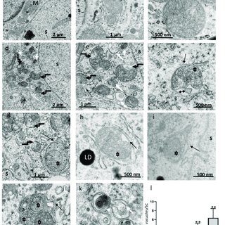
Figure 1. Ultrastructural characteristics of enhanced mitochondrial damage and mitophagy in ethanol-treated rats (ETR) Sertoli cells (SCs.) (a-c): control testes; (d-k): ETRs. Quantification of mitophagy is shown in (l). Note the normal mitochondria (M) in control testes with characteristic vesicular-type cristae. Broken black arrows (d,e,g) indicate damaged mitochondria in ETR SCs, while black arrows show autophagosomal membranes engulfing damaged mitochondria (asterisks) forming mitophagosomes. The double-head arrow indicates damaged fragmented cristae. The long and short white arrows mark autolysosomes and lysosomes, respectively. White arrow heads mark multilamellar bodies. LD: lipid droplets; S: SC nucleus. The histogram depicts quantification of mitophagic vacuoles in the control and ETRs. * p < 0.01 and ** p < 0.001 vs. control (one-way analysis of variance (ANOVA)).
Figure 2. Enhanced light chain3 (LC3)-II expression in ETR SCs with specific localization to mitophagosomes (A) immunohistochemistry (IHC) of LC3. The framed areas are magnified in the insets on the right. LC3-II puncta are marked by long thin arrows, while short thick arrows indicate SCs nuclei. The green arrow marks expression in interstitial cells. (B) Quantification of LC3 expression in control and ETR SCs. (C) Western blot of LC3. The relative expression level of the protein is normalized to actin and expressed as a fold of the control (n = 3) (this normalization applies to blots of other proteins shown below.) (D) Histogram showing significant increase of LC3-II in ETRs compared to the control. P < 0.05 (t-test). (E) LC3 immunogold labeling in the control (a) and ETR SCs Figure 2. Enhanced light chain3 (LC3)-II expression in ETR SCs with specific localization to mitophagosomes (A) immunohistochemistry (IHC) of LC3. The framed areas are magnified in the insets on the right. LC3-II puncta are marked by long thin arrows, while short thick arrows indicate SCs nuclei. The green arrow marks expression in interstitial cells. (B) Quantification of LC3 expression in control and ETR SCs. (C) Western blot of LC3. The relative expression level of the protein is normalized to actin and expressed as a fold of the control (n = 3) (this normalization applies to blots of other proteins shown below.) (D) Histogram showing significant increase of LC3-II in ETRs compared to the control. P < 0.05 (t-test). (E) LC3 immunogold labeling in the control (a) and ETR SCs (b-d). Thin arrows indicate LC3 immunogold particles, while thick arrows mark autophagosomal membranes sequestering damaged mitochondria (asterisks) forming mitophagosomes. (F) Histogram demonstrating significant increase in the number of LC3-II immunogold particles in mitophagosomes of ETRs SCs compared to the control group. P < 0.01 (t-test). M: normal mitochondria; S: SC nucleus.
Figure 5. Ultrastructural characteristics of Parkin overexpression and mitochondrial translocation in ETR SCs. (a) control; (b-d) ETRs. The long black arrows mark Parkin immunogold particles, while the short black arrows indicate damaged mitochondria. The white arrows mark the autophagosomal membrane. S: SC nucleus; M: normal mitochondria. (e) Histogram demonstrating significant increase in the number of Parkin immunogold particles in the mitochondria of ETRs SCs compared to control. ** p < 0.01 (t-test).
Figure 7. Elevated expression and colocalization of Parkin with pan cathepsin in ETRs SCs. (control, A-C), (ETRs, D-F). The framed areas in (A) and (D) are magnified in (B) and (E), respectively. Note the enhanced expression and colocalization of Parkin (red, red arrows) and pan-cathepsin (green, green arrows) upon merging (orange-yellow, orange arrows) in ETR SCs (D,E) compared to control (A,B). S: SC nucleus. DAPI (blue) was used for nuclear counterstaining. (C,F) Plot profiles demonstrating the colocalization of Parkin and pan-cathepsin defined as overlapped red and green peak at the lines positioned in images (cropped from panels B,E) just above the histograms.
Ethanol-Induced Mitochondrial Damage in Sertoli Cells is Associated with Parkin Overexpression and Activation of Mitophagy

March 2019

·

453 Reads

·

21 Citations

Cells

This study was conducted to elucidate the involvement of the PINK1-Parkin pathway in ethanol-induced mitophagy among Sertoli cells (SCs). In the research, adult rats were given intraperitoneal injections of ethanol (5 gm/kg) and sacrificed at various time periods within 24 h. Transmission electron microscopy was applied to reveal enhanced mitochondrial damage in SCs of the ethanol-treated rats (ETRs) in association with a significant increase in numbers of mitophagic vacuoles (mitophagosomes and autolysosomes) in contrast to very low levels in a control group treated with phosphate-buffered saline (PBS). This enhancement was ultra-structurally verified via observation of trapped mitochondria within LC3-labeled membranes, upregulation of LC3 protein levels, colocalization of LC3 and cytochrome c, and reduced expression of mitochondrial proteins. Importantly, Parkin expression was found to be upregulated in ETR SCs, specifically in mitochondria and mitophagosomes in addition to colocalization with PINK1 and pan-cathepsin, indicating augmented mitophagy. Transcription factor EB (TFEB, a transcription factor for autophagy and mitophagy proteins) was also found to be upregulated in nuclei of ETR SCs and associated with enhanced expression of iNOS. Enhanced Parkin-related mitophagy in ETR SCs may be a protective mechanism with therapeutic implications. To the authors' knowledge, this is the first report demonstrating the ultrastructural characteristics and molecular mechanisms of Parkin-related mitophagy in ETR SCs.


Figure 1. Ethanol-induced reduction of androgen receptor (AR) expression in ETRs testes. (A,B) Western blot analysis for AR. The relative expression level for protein was normalized to actin and expressed as fold change relative to the control (n = 3). * p < 0.01 compared to the control group. (C) Immunohistochemistry (IHC) of AR in control (a,c) and ETRs (b,d). Arrow heads indicate nuclear staining of AR in SCs, while broken arrows mark its expression in myoid cells. The black and white arrows indicate the expression of AR in Leydig cells and smooth muscles of blood vessels, respectively (red arrow: blood vessel lumen).
Figure 5. Ultrastructural features of enhanced autophagy in SCs of ETRs during stages VII-VIII. ((a,c,e): control testes; (b,d,f): ETRs). The long black arrows indicate autophagic vacuoles (AVs) containing membranous structures, while the white arrows mark damaged mitochondria. The short black arrows and arrow heads show lysosomes and lipid droplets (LDs), respectively. RS: round spermatid; S: SC nucleus; SP: sperm; M: mitochondrion.
Figure 6. Accumulation of apoptotic germ cells, retained sperm, damaged mitochondria and large LDs in SCs of ETRs at stages VII-VIII. (A) TUNEL labeling of apoptotic cells (white arrow heads) in control (a) and ETR (b) testes. The white arrows mark retained sperm with TUNEL positivity apparently phagocytosed by SCs. (B) TEM showing apoptotic germ cells (stars in c-e) and retained sperm (short arrows in e) phagocytosed by SCs of ETRs. The insets in f are higher magnifications of the boxed areas, demonstrating a mitophagosome marked by green arrow (right inset) and autolysosome marked by blue arrow head (left inset). The long black arrows indicate AVs, while the black arrow head in the right inset marks autophagosomal membrane. M: damaged mitochondria; S: SC nucleus; LD: lipid droplet.
Ethanol-Induced Autophagy in Sertoli Cells Is Specifically Marked at Androgen-Dependent Stages of the Spermatogenic Cycle: Potential Mechanisms and Implications

January 2019

·

176 Reads

·

29 Citations

International Journal of Molecular Sciences

In a recent study, we reported that acute ethanol exposure enhanced autophagy in Sertoli cells (SCs) of adult rats. However, further research is needed to clarify the specific spermatogenic stage exhibiting the highest autophagic response, the mechanisms behind such specificity, and the related relevance to sperm. This brief report provides results indicating that stages VII-VIII (androgen-dependent or spermiation stages) of the spermatogenic cycle exhibited more marked autophagic response in acute-ethanol treated rats (ETRs) than other stages based on suppression of androgen receptor (AR), analysis of microtubule-associated protein 1 light chain 3 (LC3) (an autophagosomal marker) immunostaining in SCs, double labeling of LC3 and lysosomal proteins and electron microscopy. Ultrastructural observations and TUNEL method revealed a notable presence of phagocytosed apoptotic germ cells and retained sperm in SCs of ETRs at these specific stages-a finding rarely observed in control testes. In addition, PTEN-induced putative kinase 1 (PINK1) (a sensor of mitochondrial damage and mitophagy) and giant lipid droplets were found to have accumulated in SCs of ETRs at same stages. Our data show novel findings indicating that stages VII-VIII of the spermatogenic cycle exhibit high levels of autophagy, specifically under stress conditions, as expressed by the term autophagic stages. This stage-specific upregulation of autophagy in SCs may be related to AR suppression, mitochondrial damage, lipid accumulation, and phagocytosis of apoptotic cells. The phenomenon may be an essential part of ensuring the viability of SCs and supporting germ cells in toxic environments.


Using Artificial DNA Sequence to Suppress Non-specific Bindings in Crude Nuclear Extract During Surface Plasmon Resonance Assay

September 2018

·

35 Reads

·

2 Citations

The Tokai journal of experimental and clinical medicine

Objective: Surface plasmon resonance (SPR) has been extensively used to characterize the interactions between molecules in terms of their binding specificity, affinity, and kinetics. Practical procedures, however, for measurement of the protein-DNA association in crude nuclear extract are yet to be defined. Methods: Crude nuclear extract was obtained from MCF-7 cells or recombinant estrogen receptor alpha (ERα) was used for analysis. To suppress signal from non-specific bindings in SPR assay using Biacore, DNA fragments with minimal protein binding activity were identified in a database for transcription factors and included in the study. Results: It is known that when analytes were purified transcription factors, the dissociation curves in Biacore sensorgrams exhibit exponential tendency. Based on statistical analysis, the dissociation phase between the ERα complex from crude nuclear extract and DNA oligonucleotides could be fitted exponentially. Following extrapolation of the dissociation phase, theoretical amount of bound antibodies could be estimated and compared for significant difference. Conclusion: Our procedures made SPR technique such as Biacore a practical technique for measurement of protein-DNA associations in crude nuclear extract with reproducible and reliable results.


Figure 1 of 2
Figure 2 of 2
Upregulation of autophagy and glycolysis markers in keloid hypoxic-zone fibroblasts: Morphological characteristics and implications

May 2018

·

591 Reads

·

27 Citations

Histology and Histopathology

Keloid is a fibro-proliferative skin disorder with tumor-like behavior and dependence on anaerobic glycolysis (the Warburg effect), but its exact pathogenesis is unknown. Although autophagy is widely accepted as a lysosomal pathway for cell survival and cellular homeostasis (specifically upon exposure to stressors such as hypoxia), very few studies have investigated the involvement of autophagy and related glycolytic effectors in keloidogenesis. Here the authors examined the expression and cellular localization of autophagy proteins (LC3, pan-cathepsin), glycolytic markers (LDH, MCT1, MCT4) and the transcription factor HIF isoforms in human keloid samples using immunohistochemical analysis and double-labeling immunofluorescence methods. Based on H&E staining and expression of CD31, keloids were compartmentalized into hypoxic central and normoxic marginal zones. Vimentin-expressing fibroblasts in the central zone exhibited greater autophagy than their marginal-zone counterparts, as evidenced by increased LC3 puncta formation and co-localization with lysosomal pan-cathepsin. LDH (a lactate stimulator), MCT4 (a lactate exporter) and HIF-1 α expression levels were also higher in central-zone fibroblasts. Conversely, HIF-2 α expression was upregulated in fibroblasts and endothelial cells of the peripheral zone, while MCT1 was expressed in both zones. Taken together, these observations suggest that upregulation of autophagy and glycolysis markers in keloid hypoxic-zone fibroblasts may indicate a prosurvival mechanism allowing the extrusion of lactate to marginal-zone fibroblasts via metabolic coupling. The authors believe this is the first report on differential expression of autophagic and glycolytic markers in keloid-zone fibroblasts. The study results indicate that autophagy inhibitors and MCT4 blockers may have therapeutic implications in keloid treatment.


GABAB receptor regulates proliferation in the high-grade chondrosarcoma cell line OUMS-27 via apoptotic pathways

March 2018

·

153 Reads

·

27 Citations

BMC Cancer

Background: High-grade chondrosarcoma, which has a high incidence of local recurrence and pulmonary metastasis despite surgical resection, is associated with poor prognosis. Therefore, new and effective adjuvant therapies are urgently required for this disease. Gamma-aminobutyric acid (GABA), which acts as a neurotrophic factor during nervous system development, is related to the proliferation and migration of certain cancer cells. The GABAergic system, which is composed of GABA, the GABA-synthesizing enzyme glutamic acid decarboxylase (GAD), and GABA receptors, has an important function in nerve growth and development of neural crest. Therefore, the GABAergic system may play important functional roles in the proliferation of chondrosarcoma cells, which are derived from neural crest cells. We examined the anti-tumor effects of the GABAergic system on a chondrosarcoma cell line. Methods: We evaluated the underlying mechanisms of the anti-tumor effects of the GABAergic system, such as the involvement of different signaling pathways, apoptosis, and cell cycle arrest, in the high-grade chondrosarcoma cell line OUMS-27. In addition, we performed whole-cell patch-clamp recordings for Ca2+currents and evaluated the changes in intracellular Ca2+concentration via Ca2+channels, which are related to the GABABreceptor in high-grade chondrosarcoma cells. Results: The GABABreceptor antagonist CGP had anti-tumor effects on high-grade chondrosarcoma cells in a dose-dependent manner. The activities of caspase 3 and caspase 9 were significantly elevated in CGP-treated cells compared to in untreated cells. The activity of caspase 8 did not differ significantly between untreated cells and CGP-treated cells. However, caspase 8 tended to be up-regulated in CGP-treated cells. The GABABreceptor antagonist exhibited anti-tumor effects at the G1/S cell cycle checkpoint and induced apoptosis via dual inhibition of the PI3/Akt/mTOR and MAPK signaling pathways. Furthermore, the changes in intracellular Ca2+via GABABreceptor-related Ca2+channels inhibited the proliferation of high-grade chondrosarcoma cells by inducing and modulating apoptotic pathways. Conclusions: The GABABreceptor antagonist may improve the prognosis of high-grade chondrosarcoma by exerting anti-tumor effects via different signaling pathways, apoptosis, cell cycle arrest, and Ca2+channels in high-grade chondrosarcoma cells.


Expression of delta-like 3 is downregulated by aberrant DNA methylation and histone modification in hepatocellular carcinoma

March 2018

·

25 Reads

·

15 Citations

Oncology Reports

Delta-like 3 (DLL3) is a member of the Delta/Serrate/Lag-2 family of ligands for the Notch receptor and plays a role in Notch signaling. We have previously revealed that the expression of DLL3 is silenced by aberrant DNA methylation and that overexpression of DLL3 in the HuH2 hepatocellular carcinoma (HCC) cell line induced apoptosis. In the present study, we first confirmed the methylation of DLL3 in HuH2 cells and analyzed the methylation status of the DLL3 promoter region by bisulfite sequencing. Furthermore, we investigated whether other epigenetic modifications, such as histone acetylation and histone methylation, affected the expression of DLL3. Treatment with the DNA methylation inhibitor, 5-azadeoxycytidine (5-Aza-dC) slightly reactivated DLL3 mRNA expression and bisulfite sequencing revealed that CpG sites in the DLL3 promoter region of the HuH2 cells were densely-methylated. In addition, a significant increase in the expression of DLL3 was observed when the cells were treated with 5-Aza-dC in combination with the histone deacetylase inhibitor trichostatin A. However, an inhibitor of the dimethylation of histone H3 lysine 9 (H3K9me2) or the trimethylation of histone H3 lysine 27 (H3K27me3), modifications that are associated with gene silencing, had no effect on DLL3 reactivation. In combination with the findings from our previous study, these results indicated that DLL3 expression was silenced in HCC cells by DNA methylation and was more readily affected by histone acetylation than histone methylation (H3K9me2 or H3K27me3).


Citations (78)


... The inhibition of the gastrointestinal mucosal barrier is facilitated by programmed cell death, which triggers the downstream release of caspases [10]. There is a correlation between apoptosis, autophagy, mucosal erosion, and ulceration induced by IND [11]. This is evident in the upregulated pro-apoptosis Bax protein and downregulated anti-apoptosis Bcl-2 protein within the gastric mucosa [12]. ...

Reference:

Luteolin ameliorates Indomethacin-Induced Gastric Ulcer in Rats via Activation of Nrf2/HO-1 Pathway
Indomethacin can induce cell death in rat gastric parietal cells through alteration of some apoptosis- and autophagy-associated molecules
  • Citing Article
  • September 2020

International Journal of Experimental Pathology

... La cytokine IL-24 peut être utilisée comme biomarqueur de dysfonctionnement rénal aigu. En effet, un taux élevé d'IL-24 est détecté dans le sérum et dans les urines chez les patients [300]. Et l'expression de la sous-unité IL-20Rb est augmentée dans les biopsies rénales de patients pathologiques [301]. ...

Interleukin-24 is a novel diagnostic biomarker for the severity of acute kidney injury
  • Citing Article
  • December 2019

Medical Molecular Morphology

... It can also cause a phenotype in male gametes that is comparable to apoptosis [56]. Furthermore, rats' Sertoli cells exposed to acute ethanol administration could undergo mitophagy, which is crucial in keeping Sertoli cells from apoptosis and causing these cells to become activated [58]. The findings suggest that the processes governing Sertoli cell mitophagy may have therapeutic implications for male infertility. ...

Ethanol-Induced Mitochondrial Damage in Sertoli Cells is Associated with Parkin Overexpression and Activation of Mitophagy

Cells

... Sertoli cells also perform vital functions, including fluid secretion and phagocytosis, which are essential for removing residual bodies from spermatids during spermiogenesis. This process and autophagy are crucial for Sertoli cell metabolism (Almeida et al. 2008;Schulz et al. 2010;Horibe et al. 2019). In addition, Leydig cells, located amidst loose connective tissue between seminiferous tubules, synthesize steroid hormones (Schulz et al. 2010). ...

Ethanol-Induced Autophagy in Sertoli Cells Is Specifically Marked at Androgen-Dependent Stages of the Spermatogenic Cycle: Potential Mechanisms and Implications

International Journal of Molecular Sciences

... The hypoxic zone exhibited an elevated level of hypoxia-inducible factor 1 α (HIF-1α). 37 Similar to tumors, keloids display metabolic reprogramming, including enhanced anaerobic glycolysis and weakened oxidative phosphorylation under hypoxia. Compared with normal skin, lactic acid levels were higher in keloids. ...

Upregulation of autophagy and glycolysis markers in keloid hypoxic-zone fibroblasts: Morphological characteristics and implications

Histology and Histopathology

... The interplay between DNA methylation and histone acetylation promotes the overexpression of GDNF and contributes to the development of glioblastoma 10 . Abnormal DNA methylation and histone modification downregulates DLL3 and affects cell apoptosis via the Notch signaling pathway in hepatocellular carcinoma 11 . During breast cancer tumorigenesis, the downregulation of ESR1 [estrogen receptor alpha (ERα)], which induces tumor growth and metabolic reprogramming, is influenced by changes in the acetylation of the ERα promoter region and miRNA expression profiles 12 . ...

Expression of delta-like 3 is downregulated by aberrant DNA methylation and histone modification in hepatocellular carcinoma
  • Citing Article
  • March 2018

Oncology Reports

... embolization of this artery for the treatment of recurrent hemoptysis, bronchial arteriovenous malformations in addition to surgical treatment of esophageal carcinoma [2]. In the second case, we found a thoracic duct passing ventral and medial to the right bronchial artery arising from the third posterior intercostal artery associated with ectopic common bronchial artery trunk [3,4]. In the latter papers, diagrams showing such relationships between the thoracic duct and right bronchial artery in addition to the left recurrent laryngeal nerve were shown. ...

Anomalous bronchial arteries: clinically important relationships to left recurrent laryngeal nerve and thoracic duct
  • Citing Article
  • July 2011

... Matrix-assisted laser desorption/ionization mass spectrometry imaging 31 has been shown to resolve the identity of crystals in kidney tissue col- lected from rabbits presenting with kidney toxicity (as certain specific metabolites and not the parent drug candidate or endogenous biochemical). Kidney ultra- sound imaging end points also have been described as a useful noninvasive biomarker of AKI using measure- ments of echogenicity 32 and/or blood flow with or without microbubble contrast enhancement. 33,34 One company reported using histopathology of a fine-nee- dle aspirate of the kidney as an in vivo supplemental diagnostic in nonclinical studies. ...

Quantified kidney echogenicity in mice with renal ischemia reperfusion injury: evaluation as a noninvasive biomarker of acute kidney injury
  • Citing Article
  • April 2017

Medical Molecular Morphology

... Zhu et al (60) reported that beclin 1 was a substrate of caspase-3 and the cleavage of beclin 1 may contribute to inactivation of autophagy leading to augmenta- tion of apoptosis. Beclin1 protein includes a bcl-2-homology-3 domain, which can combine with bcl-2/bcl-xl and abrogate anti-apoptotic effects (51,61). In the present study, administra- tion of L-glutathione decreased the necrosis of hepatocytes. ...

Ethanol enhances thymocyte apoptosis and autophagy in macrophages of rat thymi
  • Citing Article
  • December 2016

Histology and Histopathology