Wei Peng's research while affiliated with Fujian Provincial Cancer Hospital and other places

What is this page?


This page lists the scientific contributions of an author, who either does not have a ResearchGate profile, or has not yet added these contributions to their profile.

It was automatically created by ResearchGate to create a record of this author's body of work. We create such pages to advance our goal of creating and maintaining the most comprehensive scientific repository possible. In doing so, we process publicly available (personal) data relating to the author as a member of the scientific community.

If you're a ResearchGate member, you can follow this page to keep up with this author's work.

If you are this author, and you don't want us to display this page anymore, please let us know.

Publications (7)


FIGURE 1 | Flowchart of research design. IHC, immunohistochemistry; qRT-PCR, quantitative real-time PCR; WB, Western blotting.
FIGURE 2 | The expression and prognostic value of LDHC/LDH-C4 in LUAD were analyzed based on an online database. (A) Pan-cancer view of LDHC mRNA in the UALCAN database. Expression of LDHC in LUAD tissue based on (B) sample types (LUAD vs. normal control) and (C) clinical stages in UALCAN. (D) Pancancer review of LDH-C4 protein expression using UALCAN, and all lung cancer samples were from LUAD cases (n = 111). The plotted survival curves for (E) OS and (F) PFS were drawn by the LOGpc database. (G) HR forest map of LDHC expression and lung cancer prognosis analyzed using the LOGpc platform (combined HR = 1.1542).
FIGURE 3 | Expression of LDHC in serum and serum-derived exosomes of patients with LUAD and its correlation with clinical molecular markers. (A) Identification of extracted serum-derived exosome by transmission electron microscopy. The isolated vesicles were indicated by the red arrows. (B) Immunoblotting was used to detect the expression of exosome-related marker proteins. Cyto C (mitochondrial marker) was taken as a negative control to exclude the possible mixture of cellular contamination. PT: paracancerous tissue; PC: positive control using the extracted protein of LDHC downregulated A549 cells. The expression of (C, D) serum and (E, F) serum-derived exosomal LDHC in newly diagnosed LUAD patients and patients with different clinical stages were detected by qPCR. Serum LDHC was positively correlated with the expression of (G) Ki67, (H) CEA, (I) Pro-GRP, and (J) NSE in LUAD patients. Serum-derived exosomal LDHC expression was not correlated with (K) Ki67, but positively correlated with (L) CEA, (M) Pro-GRP, and (N) NSE.
FIGURE 4 | The value of serum and serum-derived exosomal LDHC in diagnosis, efficacy evaluation, and recurrence monitoring of LUAD. ROC curve showed the diagnostic efficacy of (A) serum and (B) exosomal LDHC in distinguishing LUAD patients from healthy controls. The expression of (C, D) serum LDHC in patients with LUAD at first visit, after treatment and relapse. (E) The efficacy of serum LDHC in predicting recurrence in patients with LUAD after treatment. Expression of (F, G) exosomal LDHC in patients with recurrent LUAD. (H) The efficacy of exosomal LDHC in predicting recurrence in patients with LUAD after treatment.
FIGURE 5 | Expression of LDH-C4 protein in LUAD and its prognostic value. (A) The expression of LDH-C4 in LUAD and adjacent tissues was detected by IHC analysis based on tissue microarray. (B) The expression score of LDH-C4 protein in LUAD patients with stage I + II was lower than that in LUAD patients with stage III + IV. (C, D) Immunoblotting was used to detect the expression of LDH-C4 in LUAD patients and matched paracancerous tissues (n = 8). (E) Patients with high expression of LDH-C4 showed poorer overall survival time. (F) The 5-year relative risk analysis based on clinicopathological parameters of LUAD patients.

+2

Cancer-Testis Antigen LDH-C4 in Tissue, Serum, and Serum-Derived Exosomes Serves as a Promising Biomarker in Lung Adenocarcinoma
  • Article
  • Full-text available

June 2022

·

19 Reads

·

2 Citations

Frontiers in Oncology

Frontiers in Oncology

Wei Peng

·

Jin Chen

·

Yanping Xiao

·

[...]

·

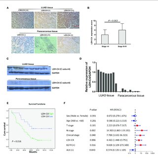
Objective As a cancer-testis antigen (CTA), human lactate dehydrogenase C4 (LDH-C4) enzyme protein encoded by the LDHC gene has been reported to be involved in the occurrence and development of various malignancies, while its expression and clinical significance in lung adenocarcinoma (LUAD) remain unclear. This study aims to investigate the expression of LDH-C4 in LUAD and its diagnostic and prognostic value.Methods The mRNA and protein levels of LDH-C4 in LUAD and adjacent normal tissues were analyzed based on the UALCAN database, and the prognostic significance was assessed using the LOGpc database. The LDHC mRNA level in serum and serum secretion of LUAD patients was determined by quantitative real-time PCR (qRT-PCR). Based on the high-throughput LUAD tissue chip combined with immunohistochemistry (IHC), the protein level of LDH-C4 in LUAD tissues was measured, and its correlation with clinicopathological features and prognosis was analyzed.ResultsLDHC expression was upregulated in LUAD, which was related to the clinical stage and poor prognosis of patients. The positive rates of LDHC mRNA expression in serum and exosome of LUAD patients were 78.3% and 66.7%, respectively. The area under the curve (AUC) of serum and exosomal LDHC in the diagnosis of LUAD was 0.8121 and 0.8925, respectively. The expression of LDHC in serum and serum-derived exosomes from LUAD patients was negatively correlated with medical treatment and positively correlated with the recurrence of LUAD. The positive expression rate of LDH-C4 in LUAD tissues was 96.7% (89/92), which was significantly higher than that in adjacent normal tissues 22.6% (19/84) (p < 0.001). The median overall survival (OS) time of patients with a high expression of LDH-C4 was significantly shorter than that of patients with low expression (34 months versus 62 months) (p = 0.016). Further relative risk analysis exhibited that the expression of LDH-C4 was an independent prognostic factor of OS in patients with LUAD.ConclusionsLDHC/LDH-C4 expression was upregulated in LUAD, and LDH-C4 could be used as a molecular indicator of the prognosis of LUAD. Serum and serum-derived exosomes of LDHC can be used as an important biomarker for the diagnosis, efficacy evaluation, and recurrence monitoring of LUAD.

Download
Share

Apelin is associated with clinicopathological parameters and prognosis in breast cancer patients

March 2022

·

17 Reads

·

8 Citations

Archives of Gynecology and Obstetrics

PurposeApelin has been shown to be a novel angiogenic factor in various cancers. However, there is limited information regarding the role of apelin in breast cancer. The aim of the present study is to examine associations between apelin, clinicopathological variables, and clinical outcome in breast cancer patients.Methods In this study, we began by investigating the apelin expression in breast cancer with long-term follow-up using immunohistochemistry. We then analyzed the relationship between apelin expression and microvessel density (MVD), lymphatic vessel density (LVD), lymph node status as well as other established clinicopathological parameters. The relationship between apelin expression and prognosis was also studied. In addition, we compared the apelin and its ligant APJ expression between 30 breast cancer samples and normal breast tissues adjacent to the breast tumors using western blot (WB) and RT-PCR.ResultsApelin protein expression was detected in the cytoplasm of the breast carcinoma cells at various intensities. Apelin expression was positive in 59.2% (84/142) of the breast cancer patients and apelin expression was significantly correlated with tumor size (p = 0.030), stage (p = 0.000), histological type (p = 0.009), MVD (p = 0.000), LVD (p = 0.000), and lymph node metastasis (p = 0.041). Survival curves determined by the Kaplan–Meier method and univariate analysis demonstrated that high expression of apelin was associated with both worse disease-free survival (p < 0.001) and overall survival (p < 0.001). Interestingly, a significant difference in apelin and APJ expression by WB as well as RT-PCR was observed between normal breast tissues adjacent to the breast tumors and breast cancer tissues.Conclusions Our results showed apelin expression was associated with tumor size, stage, histological type, MVD, LVD, lymph node metastasis and poor prognosis. The presence of apelin may be a new prognostic factor and potential therapeutic target for breast cancer.


FIGURE 1 | Tissue BATF2 downexpression in NPC tissues using IHC microarrays. (A) BATF2 is predominately expressed in the nucleus of NPC cells (× 200). (B) Elevated BATF2 expression scores are found in stage I-II NPC patients and those without recurrent tumor or metastasis in the cervical lymph nodes (CLNs). (C) Low BATF2 expression is correlated with Ki-67 upregulation in NPC tissues. (D) The Kaplan-Meier survival analysis shows that BATF2 expression is positively correlated with NPC prognosis. (E) The forest plot of risk factors for NPC prognosis (the cut-off point for HR calculation: 5-year follow-up). (F) Low BATF2 expression was detected in biopsy tissues using immunoblotting. The BATF2-overexpressed NPC cell line (CNE2) is utilized as a positive control (PC).
FIGURE 2 | Identification of serum exosomes and efficacy of serum and exosomal BATF2 expression in NPC diagnosis. (A) Morphology of serum-derived exosomes under transmission electron microscopy. The red arrows indicate isolated vesicles. (B) The immunoblotting assay shows that the exosome-specific markers, CD9 and CD63, are detectable in isolated vesicles. HC: healthy control. (C,D) Serum BATF2 mRNA expression significantly decreases in NPC versus healthy controls. (E) The ROC curve to assess the efficacy of serum BATF2 mRNA in NPC diagnosis. (F,G) Suppressed exosomal BATF2 mRNA expression in NPC patients. (H) The ROC curve to assess the efficacy of exosomal BATF2 mRNA in NPC diagnosis.
FIGURE 3 | Relationship of serum and exosomal BATF2 mRNA downregulation with clinicopathological risk factors in NPC. The correlations between serum BATF2 expression and (A) SCC-Ag, (B) CYFRA21-1, (C) CEA, and (D) EBV-DNA levels are analyzed. The downregulation of exosomal BATF2 expression is not associated with (E) SCC-Ag, (F) CYFRA21-1, (G) CEA, or (H) EBV-DNA levels in NPC patients.
FIGURE 5 | The potential mechanisms for exosomal BATF2 secretion into the circulation. (A) The process of released exosomes and shed microvesicles (SMVs) (containing BATF2 molecules) into the circulation. Early endosomal contents (proteins and nucleic acids) are either recycled to the plasma membrane (PM) or sequestered in intraluminal vesicles (ILVs), generated by the budding of the limiting membrane into the lumen of endosomes, within the larger large multivesicular bodies (MVBs). (B) Apoptotic or non-apoptotic dying cells result in the generation of apoptotic bodies (Abs). Microvesicles are shed from the blebbing PM or released from apoptotic cells. These vesicles are remnants of the degrading apoptotic cell with nuclear and cytoplasmic content (including BATF2 molecules).
Correlations between BATF2 protein expressions and clinical characteristics in NPC patients.
Diagnostic and Prognostic Potential of Circulating and Tissue BATF2 in Nasopharyngeal Carcinoma

October 2021

·

42 Reads

·

6 Citations

Frontiers in Molecular Biosciences

Background: Current biomarkers for nasopharyngeal carcinoma (NPC) are less effective for early diagnosis and prognosis. The basic leucine zipper ATF-like transcription factor 2 (BATF2) gene has been shown to have a tight association with the pathogenesis of various malignancies but received scant attention in NPC research. We aimed to assess the performances of circulating and tissue BATF2 in the diagnosis and prognosis of NPC. Materials and Methods: Immunohistochemistry (IHC) microarrays were performed to quantitate the BATF2 protein expression in NPC tissues. The relationships of BATF2 protein expression with clinicopathological characteristics and NPC prognosis were assessed. BATF2 mRNA expressions in serum and serum-derived exosomes were determined using quantitative reverse transcription-polymerase chain reaction (qRT-PCR) assay. Results: The IHC microarrays revealed a predominant nuclear expression of BATF2 in NPC cells. The Kaplan-Meier survival analysis showed that BATF2-positive NPC patients enjoyed longer overall survival than BATF2-negative patients. NPC patients with serum and exosomal BATF2 mRNA expressions made up 51.47 and 48.52% of all patients, respectively. The AUCs of serum and exosomal BATF2 mRNA expressions in discriminating NPC from healthy controls were 0.9409 and 0.8983. Patients who had received radiochemotherapy exhibited higher serum and exosomal BATF2 mRNA expressions versus the levels at baseline as well as those detected in recurrent patients. Conclusion: BATF2 is expressed cancerous tissues, serum, and serum-derived exosomes in NPC patients. Circulating and tissue BATF2 can serve as a multipurpose biomarker capable of the diagnosis, prognosis prediction, efficacy evaluation, and recurrence monitoring in NPC.


Expression and clinical implications of basic leucine zipper ATF-like transcription factor 2 in breast cancer

September 2021

·

28 Reads

·

6 Citations

BMC Cancer

Background Basic leucine zipper ATF-like transcription factor 2 (BATF2) has been reported to participate in the occurrence and development of some malignancies. Herein, we aimed to explore the expression pattern and clinical implications of BATF2 in breast cancer (BC). Methods We assessed the differences in BATF2 mRNA expression between cancerous and noncancerous tissues in BC using GEPIA and UALCAN data and in BATF2 protein expression pattern using Human Protein Atlas (HPA) data. BATF2 co-expression networks were analyzed in Coexpedia. The association between the differentially expressed BATF2 mRNA and BC prognosis was assessed using UALCAN, OSbrca, and GEPIA databases. In external validations, BATF2 protein expression in BC tissues was quantitated using a tissue microarray and immunohistochemistry (IHC) analysis, and BATF2 mRNA expression in serum and serum-derived exosomes of BC patients using real-time quantitative reverse transcription polymerase chain reaction (qRT-PCR). Results No difference in the BATF2 mRNA expression level was found between cancerous and noncancerous tissues in BC based on databases. There were low-to-moderate levels of increases in BATF2 protein expressions in BC cases from the HPA cohort. BATF2 mRNA expression was negatively correlated with androgen receptor ( AR ) and positively correlated with BRCA2 DNA repair associated (BRCA2) , marker of proliferation Ki-67 ( Mki67) , and tumor protein p53 ( TP53) expressions. Generally, BATF2 mRNA exhibited a non-significant association with BC prognosis; yet the subgroup analyses showed that triple-negative breast cancer (TNBC) patients with high BATF2 mRNA expressions had a longer overall survival (OS). Our IHC analysis revealed a positive rate of BATF2 protein expression of 46.90%, mainly located in the nucleus of cancer cells in BC, and the OS of BC patients with high BATF2 protein expressions was prolonged. The positive rates of BATF2 mRNA expressions in the serum and exosomes were 45.00 and 41.67%, respectively. Besides, the AUCs of serum and exosomal BATF2 mRNA for BC diagnosis were 0.8929 and 0.8869, respectively. Conclusions BC patients exhibit low-to-moderate expressions in BATF2 mRNA expression levels in cancerous tissues. The high BATF2 protein expression can be a potential indicator of a better BC prognosis. Serum and exosomal BATF2 mRNA levels also serve as promising noninvasive biomarkers for BC diagnosis.


Study design
a Contrast-enhanced lymphatic vessels and SonoVue-SLN; b blue-stained SLN with afferent lymph vessel
Contrast-enhanced ultrasonography and blue dye methods in detection of sentinel lymph nodes following neoadjuvant chemotherapy in initially node positive breast cancer

September 2020

·

44 Reads

·

8 Citations

Archives of Gynecology and Obstetrics

Background Recent studies show that contrast-enhanced ultrasonography (CEUS) using SonoVue has the potential to improve the performance of sentinel lymph node biopsy (SLNB) in patients with early breast cancer. However, the evidence of SLNB using CEUS in patients converting from cN1 to cN0 after neoadjuvant chemotherapy (NAC) is lacking. The aim of this prospective study is to evaluate the feasibility of CEUS using SonoVue for the identification of sentinel lymph node (SLN) and the value of the combination of CEUS and blue dye (BD) for SLNB in patients converting from cN1 to cN0 following NAC.Methods Patients with cytology-proven node positive breast cancer at the initial diagnosis (stage T1-T3N1M0) from January 2018 to January 2019, underwent NAC. Patients converting from cN1 to cN0 following NAC were enrolled and randomized into two groups for SLNB: the combination method group using CEUS and BD together, and the single BD method group. Then all patients underwent complete axillary lymph node dissection (ALND) and primary breast surgery. Compared with the final pathological results, the identification rate, sensitivity, specificity, accuracy, false negative rate, negative predictive value, positive predictive value were recorded and compared between two methods.ResultsA total of 400 patients with stage T1-T3N1M0 disease underwent NAC between January 2018 to January 2019, among which 134 (33.5%) patients had clinically negative node confirmed by imaging after NAC and randomized into two groups. Each group included 67 cases. In the combination method group, contrast-enhanced lymphatic vessels in 66 cases of 67 were clearly visualized by US soon after the periareolar injection of SonoVue and the SLNs were accurately localized. The identification rate of the combination method was 98.5%%, which was significantly higher than 83.6% (56/67) using the single BD method. The mean numbers of SLNs identified by the combination method was higher than that by the single BD method. Compared with pathological diagnosis, sensitivity, specificity, accuracy, the positive predictive value, the negative predictive value, and the FNR of the combingation method were 84.4%, 100%, 89.4%, 100%, 75%, and 15.6%, respectively. In contrast, sensitivity, specificity, accuracy, the positive predictive value, the negative predictive value, and the FNR using single blue dye were 73.9%, 100%, 89.3%, 100%, 84.6%, and 26.1%, respectively. The FNR using the combination method was significantly lower than that using single BD.Conclusion Identification of SLNs in patients converting from cN1 to cN0 following NAC by CEUS is a technically feasible. The combination of CEUS and BD is more effective than BD alone for SLNB in patients converting from cN1 to cN0 following NAC.


Fig. 1 Ultrasound image of seroma after removal of the drains in a breast cancer patient who underwent MRM on the right side
Patients and tumor characteristics
Comparison of the two treatment methods for seromas
Indwelling cannulas (16-gauge)
Drainage of seroma using indwelling cannulas in a breast cancer patient who suffered postmastectomy seroma after drainage removal
Prospective comparison of indwelling cannulas drain and needle aspiration for symptomatic seroma after mastectomy in breast cancer patients

January 2020

·

977 Reads

·

6 Citations

Archives of Gynecology and Obstetrics

Aims Postoperative seroma is the most frequent sequelae after mastectomy and axillary surgery with no optimal regimens for seroma resolution recommended in routine clinical. Indwelling cannulas with needle and catheter have been widely used in long-term medication therapies, but evidence of indwelling cannulas in seroma management after mastectomy is lacking. The purpose of this study is to evaluate the feasibility of indwelling cannulas in seroma management after mastectomy. Methods Patients who underwent modified radical mastectomy (MRM) and developed symptomatic seroma after removal of the drains between August 2017 and December 2018, were randomized into two groups either indwelling cannulas drain of seroma (Group A) or needle aspiration of seroma (Group B). We prospectively compared the number of visits for seroma, the time from removal of the drain to the final seroma resolution and the cost between the methods. Results A total of 860 patients underwent MRM between August 2017 and December 2018, among which 86 patients who developed symptomatic seroma after removal of the drains, were randomized into two groups either Group A or Group B. The number of visits for seroma in Group A was 2.35 ± 0.69 times, which was less than those in Group B (4.86 ± 1.06 times). Similarly, the time of drain removal to final seroma resolution in Group A was 4.65 ± 0.78 days, which was shorter than 7.09 ± 1.54 in Group B. In Group A, the total mean cost per patient (25.81 ± 7.71 RMB) was less than the total mean cost per patient (49.30 ± 9.85 RMB) in Group B. Cost savings were noted with using indwelling cannulas in seroma management. Conclusion It is feasible to drain indwelling cannulas drain for postmastectomy seroma, with less visits for patients, rapid seroma resolution and less cost. Indwelling cannulas can be an efficient, cost effective solution to treat symptomatic seroma after breast surgery.


Abnormally expressed microRNA as auxiliary biomarkers for nasopharyngeal carcinoma: A meta‑analysis

November 2017

·

15 Reads

·

3 Citations

Biomedical Reports

Aberrant expression of microRNA (miRNA) has been highlighted as a helpful indicator to aid in nasopharyngeal carcinoma (NPC) diagnosis. The present meta-analysis aimed to validate the efficacy of miRNA as potential biomarkers for NPC detection. Publication searches were conducted on the online PubMed and EMBASE databases from inception to June 2016. A bivariate meta-analysis was performed to generate the diagnostic parameters based on Meta-Disc 1.4 and Stata 12.0 programs. Sensitivity analysis and meta-regression tests were applied to trace heterogeneity sources among eligible studies. A total of six studies comprising 528 patients with NPC and 252 matched controls were enrolled. Results from the present meta-analysis demonstrated that miRNA testing achieved a pooled sensitivity of 0.78 [95% confidence interval (CI), 0.70-0.84] and specificity of 0.79 (95% CI, 0.73-0.84) in confirming NPC, corresponding to an area under the curve (AUC) value of 0.85. Additionally, the pooled diagnostic odds ratio was estimated to be 9.01 (95% CI, 5.62-14.44), along with a positive likelihood ratio of 2.81 (95% CI, 2.19-3.61) and negative likelihood ratio of 0.35 (95% CI, 0.28-0.44). Additionally, the stratified analyses revealed that paralleled testing of miRNA sustained a pooled accuracy superior compared with that of single miRNA testing (sensitivity, 0.88 vs. 0.70; specificity, 0.85 vs. 0.69; AUC, 0.95 vs. 0.75). Testing of miRNA harbors a moderate diagnostic efficacy and is acceptable as an auxiliary biomarker for NPC diagnosis.

Citations (6)


... Hu et al. revealed a correlation between apelin expression and various clinicopathological parameters including tumor size, stage, histological type, lymph node metastasis, and adverse prognosis in breast cancer. The identification of apelin as a potential prognostic factor suggests its utility as a novel therapeutic target in breast cancer [100]. Gourgue et al. suggested that apelin may be a major factor contributing to tumor growth and metastasis in triple-negative breast cancer in obese patients [101]. ...

Reference:

The Role of Adipokines in Tumor Progression and Its Association with Obesity
Apelin is associated with clinicopathological parameters and prognosis in breast cancer patients

Archives of Gynecology and Obstetrics

... Among the sarcoma-related risk factors, the activation of oncogenes and silencing of tumor suppressor genes play vital roles during sarcoma tumorigenesis (2,8). Recently, we and others have identified basic leucine zipper transcription factor ATF-like 2 (BATF2) as a tumor suppressor (9)(10)(11)(12)(13)(14)(15)(16)(17)(18). BATF2 is mainly expressed in normal cells but not in the corresponding tumor cells, while the restoration of BATF2 inhibits cancer cell proliferation, invasion and metastasis (13,14), highlighting its potential as a therapeutic target in cancer. ...

Diagnostic and Prognostic Potential of Circulating and Tissue BATF2 in Nasopharyngeal Carcinoma

Frontiers in Molecular Biosciences

... CD36, as a fatty acid receptor and a toll-like receptor co-receptor [32], has been reported to contribute to epithelial-tomesenchymal transition and metastasis of GC [33][34][35][36][37][38], while its role in the TME of gastric cancer remains unclear. BATF2, the basic leucine zipper ATF-like transcription factor 2 [39], is predicted to enhance DNA-binding transcription factor activity and reverse multidrug resistance in GC cells [40][41][42]. MYB is a critical transcription factor responsible for regulating hematopoiesis. ...

Expression and clinical implications of basic leucine zipper ATF-like transcription factor 2 in breast cancer

BMC Cancer

... Three of these were excluded because the treatment response of the primary tumor was assessed with CEUS [32,33,42]. Two of the others addressed the finding of a sentinel lymph node following neoadjuvant therapy in breast cancer [43] and conversion surgery in gastric cancer [44] and were therefore excluded. In the paper of McCarville et al., only one of the lesions studied was a lymph node [45]. ...

Contrast-enhanced ultrasonography and blue dye methods in detection of sentinel lymph nodes following neoadjuvant chemotherapy in initially node positive breast cancer

Archives of Gynecology and Obstetrics

... Postoperative breast seromas occur when tissue disruption and inflammation result in exudative fluid accumulation within a potential space created by the surgical procedure (1)(2)(3)(4)(5)(6)(7)(8)(9)(10)(11). Seromas increase the risk of infection, demand additional allocation of limited healthcare resources, and may delay adjuvant therapy (8,(12)(13)(14)(15). Multiple technical factors influence the risk of seroma, including the choice of dissection tools (use of cautery results in higher seroma rates versus sharp or ultrasonic dissection), the obliteration of dead space using suture techniques, the use and management of postoperative drains, and postoperative immobilization of the ipsilateral arm (1)(2)(3)(4)(5)16). ...

Prospective comparison of indwelling cannulas drain and needle aspiration for symptomatic seroma after mastectomy in breast cancer patients

Archives of Gynecology and Obstetrics

... The quality assessment of the included studies utilized the 14-item quality assessment for diagnostic accuracy studies tool (14-item QUADAS) and Newcastle-Ottawa Scale (NOS) checklist. 7,8 For studies representing data as plots or graphs, we retrieved data using GetData Graph Digitizer, v2.26 (http://getdata-graph-digitizer. com/), Engauge Digitizer 4.1 (http://digitizer.sourceforge.net/). ...

Abnormally expressed microRNA as auxiliary biomarkers for nasopharyngeal carcinoma: A meta‑analysis
  • Citing Article
  • November 2017

Biomedical Reports