Samuel J Reich's research while affiliated with University of Pennsylvania and other places

What is this page?


This page lists the scientific contributions of an author, who either does not have a ResearchGate profile, or has not yet added these contributions to their profile.

It was automatically created by ResearchGate to create a record of this author's body of work. We create such pages to advance our goal of creating and maintaining the most comprehensive scientific repository possible. In doing so, we process publicly available (personal) data relating to the author as a member of the scientific community.

If you're a ResearchGate member, you can follow this page to keep up with this author's work.

If you are this author, and you don't want us to display this page anymore, please let us know.

Publications (11)


TABLE 1 . MEAN CONCENTRATION OF RADIOACTIVITY IN OCULAR TISSUES OF DUTCH-BELTED RABBITS FOLLOWING A SINGLE INTRAVITREAL BOLUS INJECTION OF 3 H-BEVASIRANIB. Concentration of radioactivity, μg eq/g a
TABLE 4 . LEVELS OF INTACT BEVASIRANIB IN INDIVIDUAL RABBIT TISSUES FOLLOWING INTRAVITREAL INJECTION.
Ocular biodistribution of bevasiranib following a single intravitreal injection to rabbit eyes
  • Article
  • Full-text available

May 2008

·

141 Reads

·

67 Citations

Molecular Vision

Nadine S Dejneka

·

Shanhong Wan

·

Ottrina S Bond

·

[...]

·

Samuel J Reich

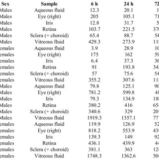
The primary objective of these investigations was to determine the ocular biodistribution of bevasiranib, a small interfering RNA (siRNA) targeting vascular endothelial growth factor A (VEGF-A), following a single intravitreal injection to rabbit eyes. A tissue distribution and pharmacokinetic study was conducted with (3)H-bevasiranib prepared in balanced-salt solution (BSS). Single doses of either 0.5 mg/eye or 2.0 mg/eye of (3)H-bevasiranib were given by intravitreal injection to Dutch-Belted rabbits (both eyes were treated). Subgroups of rabbits were serially-sacrificed at various times up to 7 days following dosing for collection of tissue samples. The right eye of each rabbit was collected whole, and the left eye was dissected to isolate five ocular tissues. All samples were analyzed by liquid scintillation counting to determine the concentrations of bevasiranib equivalents. An ocular disposition study was also performed with non-radiolabeled bevasiranib, which was administered to Dutch-Belted rabbit eyes via intravitreal injection at a dose of 2.0 mg/eye. Twenty-four hours post-dose, the eyes were enucleated and dissected into eight individual ocular structures that were analyzed for intact bevasiranib using a locked nuleic acid (LNA) noncompetitive hybridization-ligation enzyme-linked immunosorbent assay. Following intravitreal injection of 0.5 mg or 2.0 mg radiolabeled bevasiranib to Dutch-Belted rabbits, bevasiranib was detected in the vitreous, iris, retina, retinal pigment epithelium (RPE), and sclera (+choroid). As expected, the highest concentrations were found in the vitreous, and vitreous levels steadily decreased over time, while concentrations of radioactivity in the other ocular tissues increased to maximum values between 24 h and 72 h after dosing. Of these tissues, the highest concentration of radioactivity was detected in the retina. The LNA assay further confirmed the presence of intact bevasiranib in these tissues 24 h following intravitreal injection of non-radiolabeled bevasiranib (2 mg/eye). These studies demonstrate distribution of bevasiranib throughout the eye following intravitreal injection, including extensive uptake into the retina.

Download
Share

Intravitreal injection of vascular endothelial growth factor small interfering RNA inhibits growth and leakage in a nonhuman primate, laser-induced model of choroidal neovascularization

October 2004

·

36 Reads

·

131 Citations

Retina

Purpose: To determine the safety and efficacy of small interfering RNA (siRNA) directed against vascular endothelial growth factor (VEGF) in a nonhuman primate model of laser-induced choroidal neovascularization (CNV). Methods: Each animal received laser rupture of Bruch's membrane to induce CNV in both eyes. Each animal was then randomized to receive 0.05 mL of either vehicle alone or VEGF siRNA at 70 microg, 150 microg, or 350 microg in both eyes by intravitreal injection. Eyes were monitored weekly by ophthalmic examination, color photography, and fluorescein angiography for 36 days after laser injury. Electroretinograms were measured at baseline and at 5 weeks after laser. CNV on fluorescein angiograms were measured for area and graded for clinically significant leakage in a standardized, randomized, and double-masked fashion on days 15, 22, 29, and 36 after laser. Results: VEGF siRNA did not cause any change in electroretinographic, hemorrhage, inflammation, or clinical signs of toxicity. A single administration of VEGF siRNA significantly inhibited growth of CNV and attenuated angiographic leakage in a dose-dependent manner. Conclusion: Intravitreal injection of VEGF siRNA is capable of inhibiting the growth and vascular permeability of laser-induced CNV in a nonhuman primate in a dose-dependent manner. This study demonstrates preclinical proof of a principle that supports proceeding to clinical studies of VEGF siRNA in patients with exudative age-related macular degeneration.


Re: Intravitreal injection of vascular endothelial growth factor small interfering RNA inhibits growth and leakage in a nonhuman primate, laser-induced model of choroidal neovascularization

September 2004

·

20 Reads

·

141 Citations

Retina

To determine the safety and efficacy of small interfering RNA (siRNA) directed against vascular endothelial growth factor (VEGF) in a nonhuman primate model of laser-induced choroidal neovascularization (CNV). Each animal received laser rupture of Bruch's membrane to induce CNV in both eyes. Each animal was then randomized to receive 0.05 mL of either vehicle alone or VEGF siRNA at 70 microg, 150 microg, or 350 microg in both eyes by intravitreal injection. Eyes were monitored weekly by ophthalmic examination, color photography, and fluorescein angiography for 36 days after laser injury. Electroretinograms were measured at baseline and at 5 weeks after laser. CNV on fluorescein angiograms were measured for area and graded for clinically significant leakage in a standardized, randomized, and double-masked fashion on days 15, 22, 29, and 36 after laser. VEGF siRNA did not cause any change in electroretinographic, hemorrhage, inflammation, or clinical signs of toxicity. A single administration of VEGF siRNA significantly inhibited growth of CNV and attenuated angiographic leakage in a dose-dependent manner. Intravitreal injection of VEGF siRNA is capable of inhibiting the growth and vascular permeability of laser-induced CNV in a nonhuman primate in a dose-dependent manner. This study demonstrates preclinical proof of a principle that supports proceeding to clinical studies of VEGF siRNA in patients with exudative age-related macular degeneration.


FIG. 1. Fetal gene delivery and the resulting transgene expression in the retina at birth (A to C) and onset of EGFP expression following fetal delivery of AAV2/1, AAV2/2, and AAV2/5 (D to F). (A) Sub- retinal injection on E14. Following excision of the uterine wall, the fetus, which remained attached to the uterus via the placenta, was ex- posed and injected using a glass micropipette. (B) Fluorescence mi- croscopy. A transverse section of the head at birth (P0; ϫ 5 magni fi - cation) following unilateral injection of AAV2/1-CMV-EGFP on E14. (C) EGFP fl uorescence ( ϫ 20) on P0 following injection of AAV2/1- CMV-EGFP on E14. NBL, neuroblastic layer. Arrows indicate RPE. (D to F) Transgene expression was evident on E14, P3, and E16 following delivery of AAV2/1 (D), AAV2/2 (E), and AAV2/5 (F), respectively, by injection on E13. 
FIG. 2. Histological analysis of EGFP expression in adult retinas following gene delivery of AAV2/1, AAV2/2, and AAV2/5 into retinas of fetuses (A to C), newborns (D to F), and adults (readministration) (G to I). Arrows indicate M  ̈ ller cells. (A to C) Animals were injected on E14 and sacri fi ced 2 to 3 months later. (D to F) Animals were injected on P0 and sacri fi ced on P30. (G to I) Animals were injected on E14. Virus was readministered to the contralateral eye on P30. Animals were sacri fi ced on P60. ONL, outer nuclear layer; INL, inner nuclear layer; GCL, ganglion cell layer. 
FIG. 3. EGFP expression following fetal delivery of AAV2/5- CMV-EGFP. Injections were performed on E13. (A) Indirect fl uores- cent ophthalmoscopy. EGFP expression was evident throughout the retina on P90. Arrowhead, optic disk. Small arrows indicate borders of the EGFP-expressing region. (B) Histological appearance of the retina shown in panel A. 
FIG. 4. No adverse effect on visual function after fetal subretinal injection of AAV vectors. (A) ERG results for a 3-month-old mouse following fetal delivery on E13 of AAV2/5-CMV-EGFP to the treated eye. Histological analysis results for this animal are shown in Fig. 3. Traces at the top row of the graphs represent scotopic b-waves elicited by a fl ash estimated to isomerize ϳ 9 rhodopsin molecules per rod; the arrow at the left indicates the amplitude (rod b max ). The middle row shows ERGs evoked by a fl ash estimated to isomerize ϳ 100,000 rho- dopsin molecules per rod. The initial, cornea-negative de fl ection ( 2 ) represents the a-wave and the fi eld potential caused by suppression of rod dark current; the amplitude of the a-wave (rod a max ) was saturated by this fl ash intensity. The lowest row of ERGs shows the results of a saturating cone-driven b-wave response (cone b max ) elicited by an intense white fl ash in the presence of steady background light that suppressed all rod and rod-driven responses are shown. (B) Ampli- tudes (mean Ϯ standard deviations) of ERG components at 2 to 3 months from experiments with fi ve mice that received subretinal in- jections of AAV vectors during fetal development ( n ϭ 2 for AAV2/1, n ϭ for 2 AAV2/5, n ϭ for 1 AAV2/2). 
FIG. 5. Schematic representations of cells transduced in the retina following administration of AAV2/1-, AAV2/2-, and AAV2/5-CMV-EGFP. (A) Virus delivery on E13. The data for the onset of expression are shown. (B) Virus delivery at birth. (C) Virus delivery on P30 (readministration to contralateral eye). 
Delivery of Adeno-Associated Virus Vectors to the Fetal Retina: Impact of Viral Capsid Proteins on Retinal Neuronal Progenitor Transduction

July 2003

·

71 Reads

·

45 Citations

Journal of Virology

Journal of Virology

The development of fetal ocular gene transfer may be useful as a therapeutic tool for the prevention of retinal genetic disorders with congenital or early clinical manifestations. In this study we explored the neural progenitor transduction patterns of adeno-associated virus (AAV) vectors following delivery to the developing retina. Recombinant vectors with the same genome carrying the enhanced green fluorescent protein (EGFP) transgene packaged in capsids of differing serotypes (serotypes 1, 2, and 5, termed AAV2/1, AAV2/2, and AAV2/5, respectively) were created. Delivery of the AAV vectors during early retinal development resulted in efficient and stable transduction of retinal progenitors. Vector surface proteins and the developmental status of the retina profoundly affected viral tropism and transgene distribution. The procedure is not detrimental to retinal development and function and therefore provides a safe delivery vehicle for potential therapeutic applications and a means of assessing the mechanisms of retina development and disease.


Gene therapy for ocular neovascularization: A cure in sight

July 2003

·

13 Reads

·

23 Citations

Current Opinion in Genetics & Development

Recent advances in the field of ocular gene transfer have led researchers pursuing treatment for age-related macular degeneration and diabetic retinopathy to turn to gene therapy for the answer. Viral vector-mediated delivery of both anti-angiogenic proteins and molecules that inhibit the eye's endogenous pro-angiogenic factors has successfully diminished the pathology of ocular diseases in rodent models. A gene-therapy solution to the debilitating blindness caused by ocular neovascularization may be on the horizon.


Small interfering RNA (siRNA) targeting VEGF effectively inhibits ocular neovascularization in a mouse model

June 2003

·

163 Reads

·

441 Citations

Molecular Vision

RNA interference mediated by small interfering RNAs (siRNAs) is a powerful technology allowing the silencing of mamalian genes with great specificity and potency. The purpose of this study was to demonstrate the feasibility of RNA interference mediated by siRNA in retinal cells in vitro and in the murine retina in vivo. siRNAs specific for enhanced green fluorescent protein (EGFP) and murine and human vascular endothelial growth factor (VEGF) were designed. In vitro studies in human cell lines entailed modulation of endogenous VEGF levels through chemically induced hypoxia. Effects of siRNA treatment on these levels were measured by ELISA. In vivo studies evaluating effects of siRNA on levels of EGFP and VEGF were performed by co-injecting recombinant viruses carrying EGFP or hVEGF cDNAs along with the appropriate siRNAs subretinally in mice. Additional studies aimed at blocking production of endogenous mVEGF were performed using laser-induced choroidal neovascularization (CNV) in mice. Effects of in vivo treatments were evaluated ophthalmoscopically. Retinal/choroidal flat mounts were evaluated after perfusion with dextran-fluorescein. Alternatively, retinas were evaluated in histological sections or VEGF levels were measured in intact eyes using ELISA. Successful delivery of siRNA to the subretinal space was confirmed by observing significantly reduced levels of EGFP in eyes treated with Ad.CMV.EGFP plus EGFP-directed siRNA. siRNAs directed against hVEGF effectively and specifically inhibit hypoxia-induced VEGF levels in human cell lines and after adenoviral induced hVEGF transgene expression in vivo. In addition, subretinal delivery of siRNA directed against murine Vegf significantly inhibited CNV after laser photocoagulation. Delivery of siRNA can be used in vitro and in vivo to target specific RNAs and to reduce the levels of the specific protein product in the targeted cells. This work suggests that RNA interference has potential for application to studies of retinal biology and for the treatment of a variety of retinal diseases, including those involving abnormal blood vessel growth.


Efficient Trans-Splicing in the Retina Expands the Utility of Adeno-Associated Virus as a Vector for Gene Therapy

February 2003

·

47 Reads

·

92 Citations

Human Gene Therapy

Recombinant vectors based on adeno-associated virus (AAV) can efficiently transduce many different cell types, including cells of the retina, resulting in stable gene expression. A major shortcoming of this vector is its small packaging capacity. A trans-splicing approach, which reconstitutes gene expression from two independent AAV vectors, can be used to overcome the vector's packaging limitations. The efficiency of this system to date has been disappointing, and therefore its utility for therapeutic application limited. We demonstrate here that efficiency and cellular specificity of trans-splicing is dependent on selection of the appropriate AAV serotype. Efficiency of transgene expression resulting from trans-splicing in skeletal muscle approaches that obtained when delivering the intact transgene when using AAV2 vectors packaged with AAV5 capsids (AAV2/5). This expands the potential of AAV vectors for retinal gene therapy. The use of AAV2/5 also increases the efficiency of trans-splicing in photoreceptors. Selection of the appropriate AAV serotype is likely to affect efficiency of trans-splicing in other organ systems as well.


Citations (9)


... Pharmaceuticals this product has encouragingly entered phase II clinical trial for managing old age macular degeneration [40,41]. A chemically modified siRNA targeted against pain related cation channel PX23 has been intrathecally delivered into rat brain resulting in substantial clampdown of neuropathically induced pain. ...

Reference:

Bridging small interfering RNA with giant therapeutic outcomes using nanometric liposomes
Intravitreal injection of VEGF siRNA inhibits growth and leakage in a non human primate laser induced model of CNV
  • Citing Article
  • January 2004

... Vascular endothelial growth factor (VEGF), a potent and specific mitogen for vascular endothelial cells, is a critical mediator of corneal NV. Animal studies have shown that VEGF overexpression is sufficient to induce corneal NV in the eye [11,12], whereas inhibition reduces this effect [13]. Anti-VEGF has been associated with inhibition of iris and corneal NV and with suppression of the formation of new retinal vessels in primates [14][15][16]. ...

Intravitreal injection of vascular endothelial growth factor small interfering RNA inhibits growth and leakage in a nonhuman primate, laser-induced model of choroidal neovascularization
  • Citing Article
  • October 2004

Retina

... In order to determine the efficiency and tropism of different AAV serotypes toward photoreceptor cells, investigators have evaluated a variety of animal model species including but not limited to: mice and rats [10][11][12][13][14][15], pigs [9,16,17], dogs [18][19][20] and nonhuman primates [18,[21][22][23][24]. One limitation of the direct application of results from animal studies is that viral transduction can vary widely across species [25]. ...

Delivery of adeno-associated virus vectors to the fetal retina: impact of viral capsid proteins on retinal neuronal progenitor transduction.
Journal of Virology

Journal of Virology

... "Intravitreally injected naked siRNA (slightly modified, dTdT) distributed throughout the eye (vitreous, iris, retina, retinal pigment epithelium and sclera) of rabbits when administered at 2 mg/eye, and the pattern of ocular distribution was similar in male and female rabbits (Dejneka et al., 2008). The radioactive half-life (radiolabelled siRNA) in the vitreous fluid exceeded 48 h. ...

Ocular biodistribution of bevasiranib following a single intravitreal injection to rabbit eyes

Molecular Vision

... Thus, it is fundamentally different from DNA trans-splicing, in which mRNA splicing occurs in cis with the sole purpose of removing the non-coding portions between the split fragments after successful reconstitution at the genome level. Nevertheless, in many studies, dual DNA trans-splicing AAV vectors have been incompletely termed transsplicing [22][23][24] or were even mixed up with mRNA trans-splicing approaches 11 . This unfavorable terminology makes it difficult for non-experts to distinguish between the respective methods. ...

Efficient Trans-Splicing in the Retina Expands the Utility of Adeno-Associated Virus as a Vector for Gene Therapy
  • Citing Article
  • February 2003

Human Gene Therapy

... According to pharmacokinetic analysis the RNA oligonucleotide duplex could not be detected in plasma sample as rapid degradation is expected when the active substance reaches the systemic circulation. Even though RNAi technology seems to cause hardly any adverse effects in clinical trials, sequence-and target independent effects after RNA treatment cannot fully be excluded as siRNAs might induce unanticipated vascular or immune effects via the activation cell-surface toll-like receptor 3 (TLR3) so that this potential hazard of dsRNA should be taken into account for human health risk assessment (Kleinmann et al., 2008;Reich et al., 2003). ...

Small interfering RNA (siRNA) targeting VEGF effectively inhibits ocular neovascularization in a mouse model
  • Citing Article
  • June 2003

Molecular Vision

... This can be achieved using laser photocoagulation to generate burns and subsequently observing CNV formation [16,17]. These animal models have been used to explore the various molecular mechanisms of CNV and potential pharmacological implications [18,19]. The importance of VEGF signaling in the development of CNV gradually emerged [20]. ...

Re: Intravitreal injection of vascular endothelial growth factor small interfering RNA inhibits growth and leakage in a nonhuman primate, laser-induced model of choroidal neovascularization
  • Citing Article
  • September 2004

Retina

... However, this reporter also has limitations. Chief among them is that it is challenging to determine the percentage and nature of cells that are transduced in any given tissue, which would yield information that cannot be gained from measurement of the overall transgene expression (42,43). Future experiments, which are beyond the scope of the current study, with a different reporter protein (e.g., GFP) will be required to acquire such data. ...

Delivery of Adeno-Associated Virus Vectors to the Fetal Retina: Impact of Viral Capsid Proteins on Retinal Neuronal Progenitor Transduction
Journal of Virology

Journal of Virology