Petra Hermanova's research while affiliated with The Czech Academy of Sciences and other places

What is this page?


This page lists the scientific contributions of an author, who either does not have a ResearchGate profile, or has not yet added these contributions to their profile.

It was automatically created by ResearchGate to create a record of this author's body of work. We create such pages to advance our goal of creating and maintaining the most comprehensive scientific repository possible. In doing so, we process publicly available (personal) data relating to the author as a member of the scientific community.

If you're a ResearchGate member, you can follow this page to keep up with this author's work.

If you are this author, and you don't want us to display this page anymore, please let us know.

Publications (10)


FIGURE 1
FIGURE 2
FIGURE 3
FIGURE 4
FIGURE 5
The effect of oral butyrate on colonic short-chain fatty acid transporters and receptors depends on microbial status
  • Article
  • Full-text available

March 2024

·

16 Reads

Frontiers in PharmacologyFrontiers in Pharmacology
Karla Vagnerová

·

·

Martin Vodička

·

[...]

·

Jiří Pácha

Butyrate, a metabolite produced by gut bacteria, has demonstrated beneficial effects in the colon and has been used to treat inflammatory bowel diseases. However, the mechanism by which butyrate operates remains incompletely understood. Given that oral butyrate can exert either a direct impact on the gut mucosa or an indirect influence through its interaction with the gut microbiome, this study aimed to investigate three key aspects: (1) whether oral intake of butyrate modulates the expression of genes encoding short-chain fatty acid (SCFA) transporters (Slc16a1, Slc16a3, Slc16a4, Slc5a8, Abcg2) and receptors (Hcar2, Ffar2, Ffar3, Olfr78, Olfr558) in the colon, (2) the potential involvement of gut microbiota in this modulation, and (3) the impact of oral butyrate on the expression of colonic SCFA transporters and receptors during colonic inflammation. Specific pathogen-free (SPF) and germ-free (GF) mice with or without DSS-induced inflammation were provided with either water or a 0.5% sodium butyrate solution. The findings revealed that butyrate decreased the expression of Slc16a1, Slc5a8, and Hcar2 in SPF but not in GF mice, while it increased the expression of Slc16a3 in GF and the efflux pump Abcg2 in both GF and SPF animals. Moreover, the presence of microbiota was associated with the upregulation of Hcar2, Ffar2, and Ffar3 expression and the downregulation of Slc16a3. Interestingly, the challenge with DSS did not alter the expression of SCFA transporters, regardless of the presence or absence of microbiota, and the effect of butyrate on the transporter expression in SPF mice remained unaffected by DSS. The expression of SCFA receptors was only partially affected by DSS. Our results indicate that (1) consuming a relatively low concentration of butyrate can influence the expression of colonic SCFA transporters and receptors, with their expression being modulated by the gut microbiota, (2) the effect of butyrate does not appear to result from direct substrate-induced regulation but rather reflects an indirect effect associated with the gut microbiome, and (3) acute colon inflammation does not lead to significant changes in the transcriptional regulation of most SCFA transporters and receptors, with the effect of butyrate in the inflamed colon remaining intact.

Download
Share

Microbiota modulates the steroid response to acute immune stress in male mice

February 2024

·

10 Reads

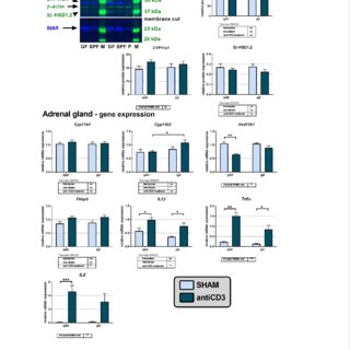
Microbiota plays a role in shaping the HPA-axis response to psychological stressors. To examine the role of microbiota in response to acute immune stressor, we stimulated the adaptive immune system by anti-CD3 antibody injection and investigated the expression of adrenal steroidogenic enzymes and profiling of plasma corticosteroids and their metabolites in specific pathogen-free (SPF) and germ-free (GF) mice. Using UHPLC-MS/MS, we showed that 4 hours after immune challenge the plasma levels of pregnenolone, progesterone, 11-deoxycorticosterone, corticosterone (CORT), 11-dehydroCORT and their 3α/β-, 5α-, and 20α-reduced metabolites were increased in SPF mice, but in their GF counterparts, only CORT was increased. Neither immune stress nor microbiota changed the mRNA and protein levels of enzymes of adrenal steroidogenesis. In contrast, immune stress resulted in downregulated expression of steroidogenic genes (Star, Cyp11a1, Hsd3b1, Hsd3b6) and upregulated expression of genes of the 3α-hydroxysteroid oxidoreductase pathway (Akr1c21, Dhrs9) in the testes of SPF mice. In the liver, immune stress downregulated the expression of genes encoding enzymes with 3β-hydroxysteroid dehydrogenase (HSD) (Hsd3b2, Hsd3b3, Hsd3b4, Hsd3b5), 3α-HSD (Akr1c14), 20α-HSD (Akr1c6, Hsd17b1, Hsd17b2) and 5α-reductase (Srd5a1) activities, except for Dhrs9, which was upregulated. In the colon, microbiota downregulated Cyp11a1 and modulated the response of Hsd11b1 and Hsd11b2 expression to immune stress. These data underline the role of microbiota in shaping the response to immune stressor. Microbiota modulates the stress-induced increase in C21 steroids, including those that are neuroactive that could play a role in alteration of HPA axis response to stress in GF animals.


Intranasal administration of EPS stimulates regulatory cellular and humoral immune responses in lung tissue. (A) Experimental design of EPS intranasal administration. BALB/c mice received intranasally 20 μg of exopolysaccharide from Lacticaseibacillus rhamnosus L900 (EPS group, n = 5) in 30 μL PBS for four consecutive days. SHAM controls (n = 5) received 30 μL PBS. (B) Histological evaluation of changes in lung tissue (magnification 200×, scale bar 100 μm). (C) Analysis of total and differential cell count in bronchoalveolar lavage (BAL). (D) Flow cytometry analysis of dendritic cells as % of CD11c⁺MHCII⁺ cells from CD45⁺ cells. (E) MHCII expression on CD11C⁺MHCII⁺ cells expressed as median fluorescence intensity (FI). (F) Percentage of FoxP3⁺ T‐cells analyzed by flow cytometry analysis and gated from CD4⁺. (G) Flow cytometry analysis of latency associated peptide (LAP, latent form of TGF‐β1)‐expressing cells gated from CD4⁺FoxP3⁺ cells. (H) Level of TGF‐β1 cytokine was determined in BAL by ELISA and expressed as pg/mL. (I) Level of TGF‐β1 cytokine was determined in supernatants of lung cell cultures by ELISA and expressed as pg/mL. (J) Percentage of IgA‐expressing B‐lymphocytes determined by flow cytometry in lung cell homogenates. (K) Level of total IgA determined in BAL by ELISA and expressed as ng/mL. (L) Levels of total IgA in cultures of lung cell homogenates. (M) Lung cells were isolated from SPF (n = 3) and germ‐free (GF) (n = 3) mice, pooled and stimulated by increasing concentration of EPS (μg/mL) for 72 h. Levels of total IgA were analyzed by ELISA and expressed as ng/mL in supernatants of cultures. All data are biological replicates expressed as mean ± standard error of mean (SEM) from one experiment. Unpaired t‐test was used for comparison between experimental groups of mice. *p < 0.05, **p < 0.01, ***p < 0.001, ****p < 0.0001.
Intranasal administration of EPS during challenge phase downregulates allergic inflammation. (A) Timeline for OVA sensitization, EPS treatment, and OVA challenge. Eight‐week‐old BALB/c mice were intraperitoneally (i.p.) sensitized with 10 μg of OVA + Alum on day 1 and 14 of experiment. Seven days after second immunization, the intranasal (i.n.) challenge with 100 μg of OVA was performed for four consecutive days. Saline (SHAM, n = 6; OVA, n = 6) or exopolysaccharide from Lacticaseibacillus rhamnosus LOCK900 (EPS/OVA, n = 6) was administered intranasally 4 h before each OVA‐challenge. (B) Total and differential cell counts were determined in bronchoalveolar lavage. (C) Representative periodic acid‐Schiff staining of lung sections from SHAM, OVA‐sensitized, and challenged mice pre‐treated with saline or EPS (scale 100 μm). Histopathological analysis of airway inflammation was performed in lung tissue. (D) Gene expression of cytokines (IL‐4, IL‐5, IL‐13, and TGF‐β1) in lung tissue was normalized using the 2−ΔCt method, and data are expressed as fold induction to SHAM control (n = 5 per group). (E) Levels of IL‐4, IL‐5, IL‐13, and TGF‐β1 cytokines were determined in bronchoalveolar lavage by ELISA (n = 6 per group). Three independent experiments were performed with similar results. All data are biological replicates expressed as mean ± standard error of mean (SEM) from one representative experiment. One‐way ANOVA with Tukey's multiple comparison test was used for comparison between experimental groups of mice. *p < 0.05, **p < 0.01, ***p < 0.001, ****p < 0.0001.
Intranasal administration of EPS activates polyspecific IgA response in Th2‐primed lung tissue. (A) Total IgA responses were determined in bronchoalveolar lavage (n = 5 per group) and (B) supernatants of lung cell cultures of mice by ELISA (n = 4 per group). (C) Number of B‐lymphocytes were analyzed in lung cell homogenates of mice by flow cytometry and shown as % from CD45⁺ cells (n = 4 per group). (D) Immunohistochemical analysis of IgA‐producing cells. Representative lung tissue cryosections (scale 40 μm) were stained with anti‐mouse IgA‐FITC antibody (green) and nuclei were counterstained by DAPI (blue). (E) IgA‐positive cells were quantified per mm² of cryosections. (F) B‐cell activating factor of the TNF family and (G) NOS2 costimulatory factors were determined by qPCR analysis in lung tissue; data were normalized using the 2−ΔCt method and expressed as fold induction compared to SHAM control (n = 5 per group). Three independent experiments were performed with similar results. All data are biological replicates expressed as mean ± standard error of mean (SEM) from one representative experiment. One‐way ANOVA with Tukey's multiple comparison test was used for comparison between experimental groups of mice. *p < 0.05, **p < 0.01, ***p < 0.001, ****p < 0.0001.
Exopolysaccharide from Lacticaseibacillus rhamnosus induces IgA production in airways and alleviates allergic airway inflammation in mouse model

May 2023

·

51 Reads

·

1 Citation

European Journal of Immunology

The currently observed high prevalence of allergic diseases has been associated with changes in microbial exposure in industrialized countries. Defined bacterial components represent a new strategy for modulating the allergic immune response. We show that intranasal administration of exopolysaccharide isolated from Lacticaseibacillus (L.) rhamnosus LOCK900 (EPS) induces TGF-β1, IgA, and regulatory FoxP3 T cells in the lungs of naive mice. Using the ovalbumin mouse model, we demonstrate that intranasal administration of EPS downregulates the development of allergic airway inflammation and the Th2 cytokine response in sensitized individuals. At the same time, EPS treatment of sensitized mice, similar to EPS-induced responses in naive mice, significantly increased the level of total, OVA-specific and also bacteria-specific IgA in bronchoalveolar lavage and the number of IgA-producing B cells in the lung tissue of these mice. Thus, EPS derived from L. rhamnosus LOCK900 can be considered a safe candidate for preventing the development of allergic symptoms in the lungs of sensitized individuals upon exposure to an allergen. This article is protected by copyright. All rights reserved.


Microbe-mediated intestinal NOD2 stimulation improves linear growth of undernourished infant mice

February 2023

·

273 Reads

·

34 Citations

Science

The intestinal microbiota is known to influence postnatal growth. We previously found that a strain of Lactiplantibacillus plantarum (strain LpWJL) buffers the adverse effects of chronic undernutrition on the growth of juvenile germ-free mice. Here, we report that LpWJL sustains the postnatal growth of malnourished conventional animals and supports both insulin-like growth factor-1 (IGF-1) and insulin production and activity. We have identified cell walls isolated from LpWJL, as well as muramyl dipeptide and mifamurtide, as sufficient cues to stimulate animal growth despite undernutrition. Further, we found that NOD2 is necessary in intestinal epithelial cells for LpWJL-mediated IGF-1 production and for postnatal growth promotion in malnourished conventional animals. These findings indicate that, coupled with renutrition, bacteria cell walls or purified NOD2 ligands have the potential to alleviate stunting.


Butyrate Treatment of DSS-Induced Ulcerative Colitis Affects the Hepatic Drug Metabolism in Mice

July 2022

·

41 Reads

·

6 Citations

The development of inflammatory bowel disease (IBD) is associated with alterations in the gut microbiota. There is currently no universal treatment for this disease, thus emphasizing the importance of developing innovative therapeutic approaches. Gut microbiome-derived metabolite butyrate with its well-known anti-inflammatory effect in the gut is a promising candidate. Due to increased intestinal permeability during IBD, butyrate may also reach the liver and influence liver physiology, including hepatic drug metabolism. To get an insight into this reason, the aim of this study was set to clarify not only the protective effects of the sodium butyrate (SB) administration on colonic inflammation but also the effects of SB on hepatic drug metabolism in experimental colitis induced by dextran sodium sulfate (DSS) in mice. It has been shown here that the butyrate pre-treatment can alleviate gut inflammation and reduce the leakiness of colonic epithelium by restoration of the assembly of tight-junction protein Zonula occludens-1 (ZO-1) in mice with DSS-induced colitis. In this article, butyrate along with inflammation has also been shown to affect the expression and enzyme activity of selected cytochromes P450 (CYPs) in the liver of mice. In this respect, CYP3A enzymes may be very sensitive to gut microbiome-targeted interventions, as significant changes in CYP3A expression and activity in response to DSS-induced colitis and/or butyrate treatment have also been observed. With regard to medications used in IBD and microbiota-targeted therapeutic approaches, it is important to deepen our knowledge of the effect of gut inflammation, and therapeutic interventions were followed concerning the ability of the organism to metabolize drugs. This gut-liver axis, mediated through inflammation as well as microbiome-derived metabolites, may affect the response to IBD therapy.


Interactions Between Gut Microbiota and Acute Restraint Stress in Peripheral Structures of the Hypothalamic–Pituitary–Adrenal Axis and the Intestine of Male Mice

November 2019

·

136 Reads

·

48 Citations

The gut microbiota play an important role in shaping brain functions and behavior, including the activity of the hypothalamus-pituitary-adrenocortical (HPA) axis. However, little is known about the effect of the microbiota on the distinct structures (hypothalamus, pituitary, and adrenals) of the HPA axis. In the present study, we analyzed the influence of the microbiota on acute restraint stress (ARS) response in the pituitary, adrenal gland, and intestine, an organ of extra-adrenal glucocorticoid synthesis. Using specific pathogen-free (SPF) and germ-free (GF) male BALB/c mice, we showed that the plasma corticosterone response to ARS was higher in GF than in SPF mice. In the pituitary, stress downregulated the expression of the gene encoding CRH receptor type 1 (Crhr1), upregulated the expression of the Fkbp5 gene regulating glucocorticoid receptor sensitivity and did not affect the expression of the proopiomelanocortin (Pomc) and glucocorticoid receptor (Gr) genes. In contrast, the microbiota downregulated the expression of pituitary Pomc and Crhr1 but had no effect on Fkbp5 and Gr. In the adrenals, the steroidogenic pathway was strongly stimulated by ARS at the level of the steroidogenic transcriptional regulator Sf-1, cholesterol transporter Star and Cyp11a1, the first enzyme of steroidogenic pathway. In contrast, the effect of the microbiota was significantly detected at the level of genes encoding steroidogenic enzymes but not at the level of Sf-1 and Star. Unlike adrenal Sf-1, the expression of the gene Lrh-1, which encodes the crucial transcriptional regulator of intestinal steroidogenesis, was modulated by the microbiota and ARS and this effect differed between the ileum and colon. The findings demonstrate that gut microbiota have an impact on the response of the pituitary, adrenals and intestine to ARS and that the interaction between stress and the microbiota during activation of glucocorticoid steroidogenesis differs between organs. The results suggest that downregulated expression of pituitary Pomc and Crhr1 in SPF animals might be an important factor in the exaggerated HPA response of GF mice to stress.


Gut microbiota metabolizes nabumetone in vitro : Consequences for its bioavailability in vivo in the rodents with altered gut microbiome

February 2019

·

71 Reads

·

13 Citations

Xenobiotica

1. The underlying microbial metabolic activity toward xenobiotics is among the least explored factors contributing to the inter-individual variability in drug response. 2. Here, we analyzed the effect of microbiota on a non-steroidal anti-inflammatory drug nabumetone. 3. First, we cultivated the drug with the selected gut commensal and probiotic bacteria under both aerobic and anaerobic conditions and analyzed its metabolites by high-performance liquid chromatography (HPLC) with UV detection. To analyze the effect of microbiota on nabumetone pharmacokinetics in vivo, we administered a single oral dose of nabumetone to rodents with intentionally altered gut microbiome - either rats treated for three days with the antibiotic imipenem or to germ-free mice. Plasma levels of its main active metabolite 6 methoxy-2-naphthylacetic acid (6-MNA) were analyzed at pre-specified time intervals using HPLC with UV/fluorescence detection. 4. We found that nabumetone is metabolized by bacteria to its non-active metabolites and that this effect is stronger under anaerobic conditions. Although in vivo, none of the pharmacokinetic parameters of 6-MNA was significantly altered, there was a clear trend towards an increase of the AUC, Cmax and t1/2 in rats with reduced microbiota and germ-free mice.


FIGURE 1 | Germ-free mice fail to develop symptoms of OVA-induced food allergy. (A) Experimental design: conventional (CV) and germ-free (GF) mice were sensitized twice within a two-week interval by intraperitoneal (i.p.) injection of 60 µg OVA in Al(OH) 3 (alum) followed by intragastric gavage (i.g.) with 15 mg OVA in PBS 8 times within 17 days (OVA groups). Age-matched control mice were injected with PBS/alum and gavaged by PBS (ctrl groups). After the last i.g. exposure, rectal temperature was assessed and mice were sacrificed by cervical dislocation. Sera and small intestine samples were collected for further analysis. Graphic art was created in freely available professional vector graphics editor Inkscape 0.92 (https://inkscape.org/). (B) The occurrence of diarrhea was monitored for 60 min after each i.g. administration in control CV (gray squares), control GF (open squares), in OVA-treated CV (black circles), and OVA-treated GF (open circles) mice. (C) The diarrhea score was assessed according to the scoring method described in Material and Methods section. (D) Representative pictures of CV/OVA and GF/OVA mice after the last i.g. OVA exposure. (E) The rectal temperature was measured 30 min after the last i.g. exposure at day 44 of the experiment in CV/ctrl (gray bars), and GF/ctrl (dashed bars) control mice, and in CV/OVA (black bars) and GF/OVA (white bars) OVA-treated mice. Difference in the temperature before and after challenge is shown. Data are plotted as mean values ± SEM. Pooled values of at least two independent experiments (CV/ctrl n = 8, CV/OVA n = 14, GF/ctrl n = 9, GF/OVA n = 11 mice per group) are shown. **P ≤ 0.01, ****P ≤ 0.0001.
FIGURE 4 | Germ-free mice have less mature mast cells and these cells are functionally impaired. (A) Messenger RNA expression of Stem cell factor (Scf) in the jejunal tissues was determined by real-time PCR. Relative expression to β-actin is shown. CV/ctrl (gray bars) n = 5, GF/ctrl (dashed bars) n = 6, CV/OVA (black bars) n = 5, GF/OVA (white bars) n = 5 mice per group. (B) Mean fluorescence intensity of CD117 was determined by flow cytometry on CD45+FcεRIα+ cells isolated from small intestine. CV (gray bars) n = 3, GF (dashed bars) n = 3 mice per group. (C) GF and CV mice were injected with degranulation-inducing compound 48/80 or PBS and edema was recorded. Data are presented as paw width after subtraction of baseline values, GF 48/80 (black circles) n = 6, CV 48/80 (open circles) n = 8, GF PBS (open squares) n = 5, CV PBS (gray squares) n = 8 mice per group. Data are plotted as mean values ± SEM. *P ≤ 0.05, **P ≤ 0.01, and ****P ≤ 0.0001.
FIGURE 5 | Colonization of germ-free mice by the conventional microbiota but not by single bacterial strain Lactobacillus plantarum WCFS1 restores sensitivity to OVA-induced food allergy. Germ-free (GF) mice were colonized by co-housing with age-matched conventional (CV) animals (exGF group) or mono-associated with the strain L. plantarum WCFS1 (Lp group). At the age of 8 weeks, all groups were submitted to OVA-sensitization and challenge, as described in Figure 1. (A) The occurrence of diarrhea during OVA treatment. (B) The diarrhea score for each group was assessed according to the scoring described in section Materials and Methods section. (C) The rectal temperature was taken 30 min after the last i.g. exposure at day 44. Difference in the temperature before and after challenge is shown. Mast cell protease-1 (MCPT-1) levels were determined in jejunal homogenates (D), and in sera (E), by ELISA. Data are plotted as mean values ± SEM. CV/OVA (n = 8), GF/OVA (n = 5) and pooled values of two independent experiments for exGF/OVA (n = 9) and Lp/OVA (n = 13) colonized mice are shown. *P ≤ 0.05, **P ≤ 0.01, ***P ≤ 0.01, ****P ≤ 0.0001.
Germ-Free Mice Exhibit Mast Cells With Impaired Functionality and Gut Homing and Do Not Develop Food Allergy

February 2019

·

400 Reads

·

41 Citations

Background: Mucosal mast cells (MC) are key players in IgE-mediated food allergy (FA). The evidence on the interaction between gut microbiota, MC and susceptibility to FA is contradictory. Objective: We tested the hypothesis that commensal bacteria are essential for MC migration to the gut and their maturation impacting the susceptibility to FA. Methods: The development and severity of FA symptoms was studied in sensitized germ-free (GF), conventional (CV), and mice mono-colonized with L. plantarum WCFS1 or co-housed with CV mice. MC were phenotypically and functionally characterized. Results: Systemic sensitization and oral challenge of GF mice with ovalbumin led to increased levels of specific IgE in serum compared to CV mice. Remarkably, despite the high levels of sensitization, GF mice did not develop diarrhea or anaphylactic hypothermia, common symptoms of FA. In the gut, GF mice expressed low levels of the MC tissue-homing markers CXCL1 and CXCL2, and harbored fewer MC which exhibited lower levels of MC protease-1 after challenge. Additionally, MC in GF mice were less mature as confirmed by flow-cytometry and their functionality was impaired as shown by reduced edema formation after injection of degranulation-provoking compound 48/80. Co-housing of GF mice with CV mice fully restored their susceptibility to develop FA. However, this did not occur when mice were mono-colonized with L. plantarum. Conclusion: Our results demonstrate that microbiota-induced maturation and gut-homing of MC is a critical step for the development of symptoms of experimental FA. This new mechanistic insight into microbiota-MC-FA axis can be exploited in the prevention and treatment of FA in humans.


FIGURE 1 | Germ-free mice fail to develop symptoms of OVA-induced food allergy. (A) Experimental design: conventional (CV) and germ-free (GF) mice were sensitized twice within a two-week interval by intraperitoneal (i.p.) injection of 60 µg OVA in Al(OH) 3 (alum) followed by intragastric gavage (i.g.) with 15 mg OVA in PBS 8 times within 17 days (OVA groups). Age-matched control mice were injected with PBS/alum and gavaged by PBS (ctrl groups). After the last i.g. exposure, rectal temperature was assessed and mice were sacrificed by cervical dislocation. Sera and small intestine samples were collected for further analysis. Graphic art was created in freely available professional vector graphics editor Inkscape 0.92 (https://inkscape.org/). (B) The occurrence of diarrhea was monitored for 60 min after each i.g. administration in control CV (gray squares), control GF (open squares), in OVA-treated CV (black circles), and OVA-treated GF (open circles) mice. (C) The diarrhea score was assessed according to the scoring method described in Material and Methods section. (D) Representative pictures of CV/OVA and GF/OVA mice after the last i.g. OVA exposure. (E) The rectal temperature was measured 30 min after the last i.g. exposure at day 44 of the experiment in CV/ctrl (gray bars), and GF/ctrl (dashed bars) control mice, and in CV/OVA (black bars) and GF/OVA (white bars) OVA-treated mice. Difference in the temperature before and after challenge is shown. Data are plotted as mean values ± SEM. Pooled values of at least two independent experiments (CV/ctrl n = 8, CV/OVA n = 14, GF/ctrl n = 9, GF/OVA n = 11 mice per group) are shown. **P ≤ 0.01, ****P ≤ 0.0001.
FIGURE 4 | Germ-free mice have less mature mast cells and these cells are functionally impaired. (A) Messenger RNA expression of Stem cell factor (Scf) in the jejunal tissues was determined by real-time PCR. Relative expression to β-actin is shown. CV/ctrl (gray bars) n = 5, GF/ctrl (dashed bars) n = 6, CV/OVA (black bars) n = 5, GF/OVA (white bars) n = 5 mice per group. (B) Mean fluorescence intensity of CD117 was determined by flow cytometry on CD45+FcεRIα+ cells isolated from small intestine. CV (gray bars) n = 3, GF (dashed bars) n = 3 mice per group. (C) GF and CV mice were injected with degranulation-inducing compound 48/80 or PBS and edema was recorded. Data are presented as paw width after subtraction of baseline values, GF 48/80 (black circles) n = 6, CV 48/80 (open circles) n = 8, GF PBS (open squares) n = 5, CV PBS (gray squares) n = 8 mice per group. Data are plotted as mean values ± SEM. *P ≤ 0.05, **P ≤ 0.01, and ****P ≤ 0.0001.
Figure 5: Colonization of germ-free mice by the conventional microbiota but not by
Germ-free mice exhibit mast cells with impaired functionality and gut homing and do not develop food allergy

August 2018

·

136 Reads

Background: Mucosal mast cells (MC) are key players in IgE-mediated food allergy (FA). The evidence on the interaction between gut microbiota, MC and susceptibility to FA is contradictory. Objective: We tested the hypothesis that commensal bacteria are essential for MC migration to the gut and their maturation impacting the susceptibility to FA. Methods: The development and severity of FA symptoms was studied in sensitized germ-free (GF), conventional (CV) and mice mono-colonized with L. plantarum WCFS1 or co-housed with CV mice. MC were phenotypically and functionally characterized. Results: Systemic sensitization and oral challenge of GF mice with ovalbumin led to increased levels of specific IgE in serum compared to CV mice. Remarkably, despite the high levels of sensitization, GF mice did not develop diarrhea or anaphylactic hypothermia, common symptoms of FA. In the gut, GF mice expressed low levels of the MC tissue-homing markers CXCL1 and CXCL2 and harbored fewer MC which exhibited lower levels of MC protease-1 after challenge. Additionally, MC in GF mice were less mature as confirmed by flow-cytometry and reduced edema formation after injection of degranulation-provoking compound 48/80. Co-housing of GF mice with CV mice fully restored their susceptibility to develop FA. However, this did not occur when GF mice were mono-colonized with L. plantarum. Conclusion: Our results demonstrate that microbiota-induced maturation and gut-homing of MC is a critical step for the development of symptoms of experimental FA. This new mechanistic insight into microbiota-MC-FA axis can be exploited in the prevention and treatment of FA in humans.

Citations (5)


... The cell walls of bacteria contain many components that are recognized by the animals for regulating innate immune responses, such as lipopolysaccharide [23][24][25] , ADP-heptose 26 , and peptidoglycan (PGN) 27,28 . PGN, a core element of all bacterial cell walls, has been identified as a key cue for promoting animal growth in mice, as demonstrated by Schwarzer et al. 29 . ...

Reference:

Bacterial peptidoglycan acts as a digestive signal mediating host adaptation to diverse food resources in C. elegans
Microbe-mediated intestinal NOD2 stimulation improves linear growth of undernourished infant mice
  • Citing Article
  • February 2023

Science

... All mice were anesthetized with isoflurane vapor and decapitated, and the colonic tissue was collected, flash-frozen in liquid nitrogen, and stored at −80°C. The present study builds upon previous research conducted by Jourova et al. (2022) and Satka et al. (2022). Their work demonstrated that administering butyrate to SPF mice for 2 weeks before subjecting them to DSS treatment helped reduce symptoms of intestinal inflammation. ...

Butyrate Treatment of DSS-Induced Ulcerative Colitis Affects the Hepatic Drug Metabolism in Mice

... The hypothalamic-pituitary-adrenal axis (HPA axis), which releases pro-inflammatory cytokines and affects several physiological systems, including the immune system and inflammatory pathways linked to the endocrine glands, plasma, and brain, is one of the most crucial parts of the gut-brain axis [56,57]. The HPA pathway (involving the hypothalamus, pituitary, and adrenals) of the gut-brain axis is an endocrine regulatory route that regulates the CNS to govern metabolic and immunological homeostasis. ...

Interactions Between Gut Microbiota and Acute Restraint Stress in Peripheral Structures of the Hypothalamic–Pituitary–Adrenal Axis and the Intestine of Male Mice

... The administration of a broad-spectrum antibiotic, imipenem to rats could inhibit the bacterial keto reductase system and formation of inactive metabolite, however, the plasma concentration of active metabolite by the liver was not affected [59]. ...

Gut microbiota metabolizes nabumetone in vitro : Consequences for its bioavailability in vivo in the rodents with altered gut microbiome

Xenobiotica

... Previous observations have suggested that germ-free mice could exhibit reduced numbers of intestinal MCs (Schwarzer et al., 2019). Because MrgprB2 neg MMCs, but not MrgprB2 + CTMCs, seemed to preferentially develop after birth and are often located in the gut lamina propria in close proximity to the colonizing microbiome, we hypothesized that the microbiota could regulate MrgprB2 neg MC development and we therefore searched for both MC populations in WT versus germ-free adult mice. ...

Germ-Free Mice Exhibit Mast Cells With Impaired Functionality and Gut Homing and Do Not Develop Food Allergy