Michael K. Rosner's research while affiliated with Washington DC VA Medical Center and other places

What is this page?


This page lists the scientific contributions of an author, who either does not have a ResearchGate profile, or has not yet added these contributions to their profile.

It was automatically created by ResearchGate to create a record of this author's body of work. We create such pages to advance our goal of creating and maintaining the most comprehensive scientific repository possible. In doing so, we process publicly available (personal) data relating to the author as a member of the scientific community.

If you're a ResearchGate member, you can follow this page to keep up with this author's work.

If you are this author, and you don't want us to display this page anymore, please let us know.

Publications (11)


879 Comparison of Perioperative and Long-Term Outcomes Following PEEK and Autologous Cranioplasty: A Single Institution Experience
  • Article

April 2024

·

1 Read

Neurosurgery

·

Peter Norman Harris

·

Khashayar Mozaffari

·

[...]

·

Michael K. Rosner

INTRODUCTION Cranioplasty is a common neurosurgical procedure to correct congenital or acquired skull defects. Three-dimensionally (3D) printed polyether-ether-ketone (PEEK) implants are a relatively novel option for cranioplasty that has gained popularity in recent years. However, there is ongoing debate with respect to this material’s efficacy and safety compared to autologous bone grafts. The purpose of this study was to offer our institution’s experience and add to the growing body of literature on the topic. METHODS A single-institution retrospective analysis of patients undergoing cranioplasties between 2016 and 2021 was performed. Patients were divided into PEEK and autologous cranioplasty cohorts. Parameters of interest included patient demographics as well as perioperative (<3 months postoperative) and long-term outcomes (>3 months postoperative). A p-value <0.05 was considered statistically significant. RESULTS A total of 31 patients met inclusion criteria (PEEK: 15, Autologous: 16). Mean age of the entire cohort was 51.7 years (range 19-81 years). Baseline demographics using the modified frailty index (mFI) revealed greater rate of comorbidities in the autologous group (p = 0.073), which was accounted for in statistical analyses. Multiple logistic regression model revealed a significantly higher rate of surgical site infection during the immediate postoperative period in the autologous cohort (31.3% vs. 0%, p = 0.011). Otherwise, perioperative and long-term complication profiles were similar between groups. Additionally, a generalized linear model demonstrated that both cohorts had similar mean hospital length of stay (LoS) (autologous: 16.1 vs. PEEK: 10.7 days). CONCLUSIONS PEEK cranioplasty implants may offer a more favorable perioperative complication profile with similar long-term complication rates and hospital LoS compared to autologous bone implants. Future studies are warranted to confirm our findings in larger series, and further examine the utility of PEEK in cranioplasty.

Share

Multicenter comparison of Chiari malformation type I presentation in children versus adults

February 2024

·

13 Reads

·

1 Citation

Journal of Neurosurgery Pediatrics

Objective: Treatment for Chiari malformation type I (CM-I) often includes surgical intervention in both pediatric and adult patients. The authors sought to investigate fundamental differences between these populations by analyzing data from pediatric and adult patients who required CM-I decompression. Methods: To better understand the presentation and surgical outcomes of both groups of patients, retrospective data from 170 adults and 153 pediatric patients (2000-2019) at six institutions were analyzed. Results: The adult CM-I patient population requiring surgical intervention had a greater proportion of female patients than the pediatric population (p < 0.0001). Radiographic findings at initial clinical presentation showed a significantly greater incidence of syringomyelia (p < 0.0001) and scoliosis (p < 0.0001) in pediatric patients compared with adult patients with CM-I. However, presenting signs and symptoms such as headaches (p < 0.0001), ocular findings (p = 0.0147), and bulbar symptoms (p = 0.0057) were more common in the adult group. After suboccipital decompression procedures, 94.4% of pediatric patients reported symptomatic relief compared with 75% of adults with CM-I (p < 0.0001). Conclusions: Here, the authors present the first retrospective evaluation comparing adult and pediatric patients who underwent CM-I decompression. Their analysis reveals that pediatric and adult patients significantly differ in terms of demographics, radiographic findings, presentation of symptoms, surgical indications, and outcomes. These findings may indicate different clinical conditions or a distinct progression of the natural history of this complex disease process within each population, which will require prospective studies to better elucidate.


( ) Maximum intensity projection of coronal computed tomographic myelogram demonstrating diastematomyelia of the lower thoracic spinal cord as well as expansile intradural multiloculated cyst, likely intramedullary in origin arising from the right hemicord and does not fill with contrast. ( ) Intraoperative picture of intramedullary cyst. ( ) Axial T2 magnetic resonance imaging (MRI) demonstrating compression of the two spinal cords, with right (gray arrows) more compressed than left (white arrows). ( ) Sagittal T2 MRI of the spine showing compression of the left hemicord by the cyst and causing syrinx.
An Unusual Case of Neurenteric Cyst in a Patient with Split Cord Malformation
  • Article
  • Full-text available

March 2023

·

23 Reads

Georg Thieme Verlag KG

Neurenteric cyst in a split cord malformation is a rare finding. We report an adult female becoming acutely symptomatic secondary to an expanding neurenteric cyst, though previous imaging had demonstrated stability. We discuss our workup and management with surgical resection and possible etiologies of her acute decline.

Download

Impact of Extent of Resection and Adjuvant Therapy in Diffuse Gliomas of the Spine

February 2023

·

23 Reads

·

2 Citations

The Spine Journal

Background content: Diffuse gliomas of the spine (DGS)-consisting of intradural intramedullary glioblastoma, astrocytoma, and oligodendroglioma-are exceedingly rare tumors that account for about 2% of primary spinal cord tumors. Much is unknown about their optimal treatment regimen due to a relative lack of clinical outcome data. Purpose: To provide an updated analysis on treatment and outcomes in DGS. Study design/setting: Observational cohort study using The National Cancer Database (NCDB), a multicenter prospectively collected oncology outcomes database. A systematic literature review was also performed to compare the resulting data to previous series. Patient sample: Patients with histologically confirmed DGS from 2004 to 2018. Outcome measures: Long-term overall survival and short-term thirty/ninety-day post-surgical mortality, thirty-day readmission, and prolonged hospital length of stay. Methods: Impact of extent of resection and adjuvant therapy on overall survival was evaluated using Kaplan-Meier estimates and multivariable Cox proportional hazards regression. Univariate and multivariate logistic regression was used to analyze covariables and their prognostic impact on short-term surgical outcomes. Results: Of the 747 cases that met inclusion criteria, there were 439 astrocytomas, 14 oligodendrogliomas, and 208 glioblastomas. Sixty percent (n=442) of patients received radiation, and 45% (n=324) received chemotherapy. Tumor histology significantly impacted survival; glioblastoma had the poorest survival (median survival time [MS]: 12.3 months), followed by astrocytoma (MS: 70.8 months) and oligodendroglioma (MS: 71.6 months) (p<0.001). Gross total resection (GTR) independently conferred a survival benefit in patients with glioblastoma (hazard ratio [HR]: 0.194, p<0.001) and other WHO grade 4 tumors (HR: 0.223, p=0.003). Adjuvant chemotherapy also improved survival in patients with glioblastoma (HR: 0.244, p=0.007) and WHO grade 4 tumors (HR: 0.252, p<0.001). Systematic literature review identified 14 prior studies with a combined DGS mortality rate of 1.3%, which is lower than the 4% real-world outcomes calculated from the NCDB. This difference may be explained by selection biases in previously published literature in which only centers with favorable outcomes publish their results. Conclusions: There remains a paucity of data regarding treatment paradigms and outcomes for DGS. Our analysis, the largest to date, demonstrates that GTR and adjuvant therapy independently improve survival for certain high-grade subgroups of DGS. This best-available data informs optimal management for such patients.


Sugammadex and blood loss during cervical spine fusion surgery

October 2022

·

2 Reads

Journal of Anaesthesiology Clinical Pharmacology

Background and Aims Sugammadex (SUG) has been associated with changes in coagulation studies. Most reports have concluded a lack of clinical significance based on surgical blood loss with SUG use at the end of surgery. Previous reports have not measured its use intraoperatively during ongoing blood loss. Our hypothesis was that the use of SUG intraoperatively may increase bleeding. Material and Methods This was a single site retrospective study. Inclusion criteria were patients undergoing a primary posterior cervical spine fusion, aged over 18 years, between July 2015 and June 2021. The primary outcomes compared were intraoperative estimated blood loss (EBL) and postoperative drain output (PDO) between patients receiving SUG, neostigmine (NEO) and no NMB reversal agent. The objective was to determine if there was a difference in primary endpoints between patients administered SUG, NEO or no paralytic reversal agent. Primary endpoints were compared using analysis of variance with a P value of 0.05 used to determine statistical significance. Groups were compared using the Chi-squared test, rank sum or student’s t test. A logistic regression model was constructed to account for differences between the groups. Results There was no difference in median EBL or PDO between groups. The use of SUG was not associated with an increase in odds for >500 milliliters (ml) of EBL. Increasing duration of surgery and chronic kidney disease were both associated with an increased risk for EBL >500 ml. Conclusion Intraoperative use of SUG was not associated with increased bleeding. Any coagulation laboratory abnormalities previously noted did not appear to have an associated clinical significance.


FIGURE 1. A, Sagittal computed tomography and B, 3-dimensional reconstructed images of the extreme displacement and distortion of the patient's hardware and vertebral column anatomy.
FIGURE 2. An augmented reality-generated 3-dimensional model of the patient's spondyloptosis. The existing hardware has been defined as a region of interest by the neurosurgeon and then contoured by the technologist.
FIGURE 3. The exposed step-off (yellow arrows) is shown by the suction resting on the caudal lamina of the spondyloptosis deformity.
FIGURE 4. A-D, Actual intraoperative stills of the augmented reality 3-dimensional model in various planes assisting in the localization of rod breaks (A, C, and D, white arrows), ossified screw heads (D, red arrows), and sublaminar hooks (A, B, and D, yellow arrows).
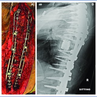
FIGURE 5. A, Intraoperative picture and B, postoperative lateral x-ray demonstrating satisfactory reduction and instrumentation of the previous spondyloptosis.
Practical Use of Augmented Reality Modeling to Guide Revision Spine Surgery: An Illustrative Case of Hardware Failure and Overriding Spondyloptosis

September 2022

·

37 Reads

·

1 Citation

Operative Neurosurgery

Background and importance: Augmented reality (AR) is a novel technology with broadening applications to neurosurgery. In deformity spine surgery, it has been primarily directed to the more precise placement of pedicle screws. However, AR may also be used to generate high fidelity three-dimensional (3D) spine models for cases of advanced deformity with existing instrumentation. We present a case in which an AR-generated 3D model was used to facilitate and expedite the removal of embedded instrumentation and guide the reduction of an overriding spondyloptotic deformity. Clinical presentation: A young adult with a remote history of a motor vehicle accident treated with long-segment posterior spinal stabilization presented with increasing back pain and difficulty sitting upright in a wheelchair. Imaging revealed pseudoarthrosis with multiple rod fractures resulting in an overriding spondyloptosis of T6 on T9. An AR-generated 3D model was useful in the intraoperative localization of rod breaks and other extensively embedded instrumentation. Real-time model thresholding expedited the safe explanation of the defunct system and correction of the spondyloptosis deformity. Conclusion: An AR-generated 3D model proved instrumental in a revision case of hardware failure and high-grade spinal deformity.


317 Comparison of One-Day Combined Versus Staged Anterior and Posterior Cervical Decompression, Fixation and Fusion

April 2022

·

9 Reads

Neurosurgery

INTRODUCTION Current management of cervical stenosis at times can involve both anterior and posterior decompression, stabilization and fusion. Although these procedures can be done in a combined or staged fashion, there is a scarcity of literature on this topic. METHODS A retrospective cohort analysis was conducted on consecutive patients at a single institution between July 2015 and April 2019 who underwent either single day combined or separate day staged surgeries. Demographics, comorbidities, length of stay, perioperative complications, and inpatient medication use were compared between cohorts. Multivariable analysis was used to adjust for differences in demographics and other confounding covariates. RESULTS 80 patients were analyzed (combined = 58, staged = 22). Comparing combined versus staged, there were no differences in baseline demographics. The administration of morphine equivalents was similar comparing combined and staged (569 vs 681 morphine equivalents, P = 0.66). Staged patients had significantly longer time in the operating room (7.2 vs 8.5 hours, P < 0.01) and longer duration of general anesthesia (6.7 vs 7.6 hours, P < 0.01). Incidence of post-operation delirium was significantly higher in the staged group (13.2% vs 50.0%), with an adjusted odds ratio of 23.3 (95% CI: 2.6 – 212.1; P < 0.01). Dysphagia was the most common post-operative complication seen in 43 patients (53.7%). Patients age > 60 had total length of stay 39.1% ± 15.0% longer than those age = 60 (multivariable adjusted mean ± standard error; P = 0.02). CONCLUSION Staging anterior and posterior cervical decompression, stabilization and fusion is associated with longer usage of the operating room, longer duration of general anesthesia, and increased incidence of perioperative complications.


Comparison of One-day Combined versus Staged Anterior and Posterior Cervical Decompression, Fixation, and Fusion

December 2021

·

18 Reads

Turkish Neurosurgery

Aim: Certain patients with cervical spinal stenosis have spinal compression both anteriorly and posteriorly, and a subset of these patients requires circumferential decompression. Surgical management consists of either combined or staged decompression; however, there is a scarcity of literature on this topic. The aim of this study was to compare the perioperative outcomes between combined and staged decompressions. Material and methods: A retrospective cohort analysis was conducted on consecutive patients admitted at a single institution between July 2015 and April 2019, who underwent either single-day combined or separate-day staged surgeries during the same hospitalization period. Demographics, comorbidities, hospital length of stay, and perioperative complications were compared between the patient groups. Results: Eighty patients (combined surgery: n=68, staged surgery: n=12) were included. Dysphagia was the most commonly reported postoperative complication in 44/80 patients (55%). There were no significant differences in the baseline demographics between the two groups. The staged surgery group had significantly longer total time in the operating room (7.2 vs. 8.5 hours, p=0.002), longer duration of general anesthesia (6.7 vs. 7.6 hours, p=0.006), and higher incidence of postoperative delirium (12.1% vs. 50% p=0.005) than the combined surgery group. The mean hospital length of stay was similar in the two groups (combined surgery: 7.5 days vs. staged surgery: 15.1 days, p=0.09). Conclusion: Staged anterior and posterior cervical decompressions, stabilizations, and fusions are associated with longer total time in the operating room, longer duration of general anesthesia, and higher incidence of postoperative delirium than combined surgeries.


Citations (3)


... TO THE EDITOR: I read with special interest the paper by Mortazavi et al. 1 about the clinical, radiographic, and treatment-related differences between pediatric and adult patients with Chiari malformation type I (Mortazavi A, Almeida ND, Hofmann K, et al. Multicenter comparison of Chiari malformation type I presentation in children versus adults. ...

Reference:

Letter to the Editor. Chiari malformation type I in adults and children
Multicenter comparison of Chiari malformation type I presentation in children versus adults
  • Citing Article
  • February 2024

Journal of Neurosurgery Pediatrics

... Standard treatment paradigms for SCGs generally mirror their intracranial counterparts, consisting of surgical resection with adjuvant radiotherapy and chemotherapy [2−4]. A population-based study reported overall survival rates at 1, 5, and 10 years were 97%, 90%, and 81% for spinal ependymomas [5], while another population-based study reported median survivals of 12.3 months for glioblastomas, 70.8 months for astrocytomas, and 71.6 months for oligodendrogliomas [2]. ...

Impact of Extent of Resection and Adjuvant Therapy in Diffuse Gliomas of the Spine
  • Citing Article
  • February 2023

The Spine Journal

... 42 There are risk factors associated with the occurrence of progressive kyphosis and neurological complications after vertebral augmentation, including location of fracture at the thoracolumbar junction, fracture type of intravertebral cleft, or wedge-type fracture, and material of nonintegration properties injected into the fractured vertebra. 42,52 Additionally, significant associations were found between cement distribution patterns and progressive kyphosis in cemented vertebrae, which affected the clinical outcome in patients after vertebral augmentation. The cement distribution included uninterlocked solid pattern, discontiguous trabecular pattern, and solid lump cement pattern. ...

Practical Use of Augmented Reality Modeling to Guide Revision Spine Surgery: An Illustrative Case of Hardware Failure and Overriding Spondyloptosis

Operative Neurosurgery