Lawrence Shapiro's research while affiliated with Columbia University and other places

What is this page?


This page lists the scientific contributions of an author, who either does not have a ResearchGate profile, or has not yet added these contributions to their profile.

It was automatically created by ResearchGate to create a record of this author's body of work. We create such pages to advance our goal of creating and maintaining the most comprehensive scientific repository possible. In doing so, we process publicly available (personal) data relating to the author as a member of the scientific community.

If you're a ResearchGate member, you can follow this page to keep up with this author's work.

If you are this author, and you don't want us to display this page anymore, please let us know.

Publications (403)


BG505 DS‐SOSIP immunized llama develops broadly neutralizing serum responses. A) Immunization schema in a llama. The llama was immunized with BG505 DS‐SOSIP subcutaneously once every 21 days (except the last boost) as indicated above the arrow line, and serum was collected 10 days post each immunization starting from day 52 as indicated beneath the arrow line. B) Antibody binding response to HIV Env probes in the immunized llama as determined by ELISA. C) Llama sera neutralization response on a 14‐strain panel. Color shading represents potency as indicated on the right of the table. D) Dendrograms of the neutralization activity of day 188 and day 271 sera on a 60‐strain panel. Dendrograms display the diversity of tested viral strains, with branches colored according to neutralization potency (non‐neutralized branches shown in gray).
Identification of neutralizing nanobodies from BG505 DS‐SOSIP immunized llama. A) Nanobody phage library construction and screening. The four probes used for phage screening are: Env Trimer (BG505 DS‐SOSIP), Fusion peptide, Glycan base trimer (BG505 DS‐SOSIP.4mut_N502‐660), and RSC3. B) Summary of epitopes of 151 nanobodies from day 188 library. 30 nanobodies selected for small panel neutralization test are grouped into 4 categories: Non‐neutralizer (one line); Weak neutralizer (two lines); Moderate neutralizer (three lines); Broad neutralizer (four lines plus one red arrow). Control nanobody J3 is marked with four lines and one blue arrow. C) Phylogenetic tree of three selected nanobody lineages. 42 nanobodies from the three lineages were tested on a 10‐strain panel first, then the top 6 candidates were further tested on an additional 15‐strain panel. Neutralization breadth of the top 6 nanobodies on a 25‐strain panel is shown beneath the nanobody names. Scale bars indicate the distance of 16.67 nucleotides (nt) in each tree. D) 25‐strain neutralization of the top 6 nanobodies from Figure S3C (Supporting Information). The broadest (G36) and most potent (R27) nanobodies were selected for further analysis.
Immunization‐elicited nanobodies, G36 and R27, in nanobody x3‐IgG2a format, show broad and potent HIV‐1 neutralization. A) 25‐strain neutralization of nanobody x3‐IgG2a. B) 208‐strain panel neutralization of G36×3‐IgG2a and R27×3‐IgG2a. Dendrograms display the diversity of tested viral strains, with branches colored according to neutralization potency (non‐neutralized branches shown in gray). IC50 shown is geometric mean. C) Comparison of neutralization breadth and potency for G36×3‐IgG2a and R27×3‐IgG2a with other human antibodies and vaccine‐elicited NHP antibodies on 208‐strain panel.
Cryo‐EM structures of nanobodies R27 and G36 in complex with HIV‐1 Env trimer reveal modes of recognition similar to J3. A) Cryo‐EM structure of nanobody R27 in complex with HIV‐1 BG505 DS‐SOSIP Env. Overall cryo‐EM density map and refined model are shown in two views with gp120 protomers colored green, cyan, and slate, respectively. The density and model of nanobody R27 is colored orange. The contour level of Cryo‐EM map is 9.5 σ. B) Cryo‐EM structure of VHH G36 in complex with HIV‐1 BG505 DS‐SOSIP Env. Overall cryo‐EM density map and refined model are shown in two views with gp120 protomers colored green, cyan, and slate, respectively. The density and model of G36 is colored magenta. The contour level of Cryo‐EM map is 9.6 σ. C) Epitopes of R27 and G36 on BG505 DS‐SOSIP. Epitopes of R27, G36, and J3 are shown in orange, magenta, and pink surfaces, respectively. R27 has a much smaller contact area on the neighboring protomer. D) Comparison of binding modes and angles. (Left) Structures of nanobodies R27, G36, and J3 are aligned with CD4 by the gp120 domain shown in green. R27, G36, and J3 are roughly in a similar position with N termini (labeled with “N”) in close proximity. (Right) The axes of R27, G36, and J3 are shown in orange, magenta, and pink rods. Axes of CD4 domain 1 and Fv domain of VRC01‐class antibody N6 are shown in yellow and olive rods for comparison. E) Detailed interactions between nanobodies and BG505 DS‐SOSIP. Residues that form hydrogen bonds and salt bridges are highlighted with sticks representation with bonds between atoms shown in gray dotted lines. Nanobodies and protomers of HIV Env are colored the same as in panels A and B. F) Alignment of nanobody sequences. Paratope residues are colored in orange and magenta, respectively. Residues interacting with neighboring protomer are colored in lighter shades. Residues interacting with both protomers are underlined. G) Nanobody mimicry of CD4 Phe43 interaction with gp120. G36, like J3, inserts Tyr99 into the “Phe43 pocket” on gp120, whereas R27 has an Ala at this position.
Ultra‐potent HIV‐1 bispecific antibodies from attaching nanobodies to the light chain of V2‐apex‐directed antibody CAP256V2LS. A) Schematic of CAP256L‐nanobody chimeras. B) 38‐strain neutralization of nanobodies. C) 80‐strain panel neutralization. Dendrograms display the diversity of tested viral strains, with branches colored according to neutralization potency (non‐neutralized branches shown in gray). D) Comparison of neutralization breadth and potency for R27 and G36 constructs with other potent antibodies on the 208‐strain panel. Data for CAP256L‐G36×3LS and CAP256L‐R27×3LS are estimated from 80‐strain data in panel C. Bispecific and trispecific antibodies are shown as stars.

+2

Ultrapotent Broadly Neutralizing Human‐llama Bispecific Antibodies against HIV‐1
  • Article
  • Full-text available

May 2024

·

34 Reads

Advanced ScienceAdvanced Science
Jianliang Xu

·

·

·

[...]

·

Peter D. Kwong

Broadly neutralizing antibodies are proposed as therapeutic and prophylactic agents against HIV‐1, but their potency and breadth are less than optimal. This study describes the immunization of a llama with the prefusion‐stabilized HIV‐1 envelope (Env) trimer, BG505 DS‐SOSIP, and the identification and improvement of potent neutralizing nanobodies recognizing the CD4‐binding site (CD4bs) of vulnerability. Two of the vaccine‐elicited CD4bs‐targeting nanobodies, G36 and R27, when engineered into a triple tandem format with llama IgG2a‐hinge region and human IgG1‐constant region (G36×3‐IgG2a and R27×3‐IgG2a), neutralized 96% of a multiclade 208‐strain panel at geometric mean IC80s of 0.314 and 0.033 µg mL⁻¹, respectively. Cryo‐EM structures of these nanobodies in complex with Env trimer revealed the two nanobodies to neutralize HIV‐1 by mimicking the recognition of the CD4 receptor. To enhance their neutralizing potency and breadth, nanobodies are linked to the light chain of the V2‐apex‐targeting broadly neutralizing antibody, CAP256V2LS. The resultant human‐llama bispecific antibody CAP256L‐R27×3LS exhibited ultrapotent neutralization and breadth exceeding other published HIV‐1 broadly neutralizing antibodies, with pharmacokinetics determined in FcRn‐Fc mice similar to the parent CAP256V2LS. Vaccine‐elicited llama nanobodies, when combined with V2‐apex broadly neutralizing antibodies, may therefore be able to fulfill anti‐HIV‐1 therapeutic and prophylactic clinical goals.

Download
Share

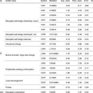
Summary of retrospective FEP+ results compared to experimental data for each system in the benchmark
Manual classification of outlier cases with absolute error > 2 kcal/mol relative to experiment. Cases within each outlier class are sorted by decreasing absolute error. All ΔΔG and error values are given in kcal/mol.
Summary of automatic flagging of probable outlier cases by magnitude of FEP+ predicted binding ΔΔG error vs. experiment.
Robust prediction of relative binding energies for protein-protein complex mutations using free energy perturbation calculations

April 2024

·

30 Reads

Computational free energy-based methods have the potential to significantly improve throughput and decrease costs of protein design efforts. Such methods must reach a high level of reliability, accuracy, and automation to be effectively deployed in practical industrial settings in a way that impacts protein design projects. Here, we present a benchmark study for the calculation of relative changes in protein-protein binding affinity for single point mutations across a variety of systems from the literature, using free energy perturbation (FEP+) calculations. We describe a method for robust treatment of alternate protonation states for titratable amino acids, which yields improved correlation with and reduced error compared to experimental binding free energies. Following careful analysis of the largest outlier cases in our dataset, we assess limitations of the default FEP+ protocols and introduce an automated script which identifies probable outlier cases that may require additional scrutiny and calculates an empirical correction for a subset of charge-related outliers. Through a series of three additional case study systems, we discuss how protein FEP+ can be applied to real-world protein design projects, and suggest areas of further study. Graphical Abstract Highlights Reliable calculation of relative binding free energy changes for most protein mutations to within ∼1 kcal/mol. Automated Protein FEP+ Groups treatment of alternate protonation states for titratable residues. Application of FEP+ methodology to “real-world” protein design projects.


Cis inhibition of co-expressed DIPs and Dprs shapes neural development

March 2024

·

4 Reads

In Drosophila, two interacting adhesion protein families, Dprs and DIPs, coordinate the assembly of neural networks. While intercellular DIP/Dpr interactions have been well characterized, DIPs and Dprs are often co-expressed within the same cells, raising the question as to whether they also interact in cis. We show, in cultured cells and in vivo, that DIP-α and DIP-δ can interact in cis with their ligands, Dpr6/10 and Dpr12, respectively. When co-expressed in cis with their cognate partners, these Dprs regulate the extent of trans binding through competitive cis interactions. We demonstrate the neurodevelopmental effects of cis inhibition in fly motor neurons and in the mushroom body. We further show that a long disordered region of DIP-α at the C-terminus is required for cis but not trans interactions, likely because it alleviates geometric constraints on cis binding. Thus, the balance between cis and trans interactions plays a role in controlling neural development.


Figures
Astrocyte morphogenesis requires self-recognition

February 2024

·

94 Reads

Self-recognition is a fundamental cellular process across evolution and forms the basis of neuronal self-avoidance1-4. Clustered protocadherins (Pcdh), comprising a large family of isoform-specific homophilic recognition molecules, play a pivotal role in neuronal self-avoidance required for mammalian brain development5-7. The probabilistic expression of different Pcdh isoforms confers unique identities upon neurons and forms the basis for neuronal processes to discriminate between self and non-self5,6,8. Whether this self-recognition mechanism exists in astrocytes, the other predominant cell type of the brain, remains unknown. Here, we report that a specific isoform in the Pcdhγ cluster, γC3, is highly enriched in human and murine astrocytes. Through genetic manipulation, we demonstrate that γC3 acts autonomously to regulate astrocyte morphogenesis in the mouse visual cortex. To determine if γC3 proteins act by promoting recognition between processes of the same astrocyte, we generated pairs of γC3 chimeric proteins capable of heterophilic binding to each other, but incapable of homophilic binding. Co-expressing complementary heterophilic binding isoform pairs in the same γC3 null astrocyte restored normal morphology. By contrast, chimeric γC3 proteins individually expressed in single γC3 null mutant astrocytes did not. These data establish that self-recognition is essential for astrocyte development in the mammalian brain and that, by contrast to neuronal self-recognition, a single Pcdh isoform is both necessary and sufficient for this process.


Genetic composition, epitope, and neutralization function of H7.HK mAbs
Allosteric Neutralization by Human H7N9 Antibodies

October 2023

·

25 Reads

The avian influenza A virus H7N9 causes severe human infections with more than 30% fatality despite the use of neuraminidase inhibitors. Currently there is no H7N9-specific prevention or treatment for humans. From a 2013 H7N9 convalescent case occurred in Hong Kong, we isolated four H7 hemagglutinin (HA)-reactive monoclonal antibodies (mAbs) by single B cell cloning, with three mAbs directed to the HA globular head domain (HA1) and one to the HA stem region (HA2). Two clonally related HA1-directed mAbs, H7.HK1 and H7.HK2, potently neutralized H7N9 and protected mice from a lethal H7N9/AH1 challenge. Cryo-EM structures revealed that H7.HK1 and H7.HK2 bind to a β14-centered surface partially overlapping with the antigenic site D of HA1 and disrupt the 220-loop that makes hydrophobic contacts with sialic acid on the adjacent protomer, thus affectively blocking viral entry. The more potent mAb H7.HK2 retained full HA1 binding and neutralization capacity to later H7N9 isolates from 2016-2017, which is consistent with structural data showing that the antigenic mutations of 2016-2017 from the 2013 H7N9 only occurred at the periphery of the mAb epitope. The HA2-directed mAb H7.HK4 lacked neutralizing activity but protected mice from the lethal H7N9/AH1 challenge when engineered to mouse IgG2a enabling Fc effector function in mice. Used in combination with H7.HK2 at a suboptimal dose, H7.HK4 augmented mouse protection. Our data demonstrated an allosteric mechanism of mAb neutralization and augmented protection against H7N9 when a HA1-directed neutralizing mAb and a HA2-directed non-neutralizing mAb were combined.


Structure-based design of a single-chain triple-disulfide-stabilized fusion-glycoprotein trimer that elicits high-titer neutralizing responses against human metapneumovirus

September 2023

·

50 Reads

·

2 Citations

The Pneumoviridae family of viruses includes human metapneumovirus (HMPV) and respiratory syncytial virus (RSV). The closely related Paramyxoviridae family includes parainfluenza viruses (PIVs). These three viral pathogens cause acute respiratory tract infections with substantial disease burden in the young, the elderly, and the immune-compromised. While promising subunit vaccines are being developed with prefusion-stabilized forms of the fusion glycoproteins (Fs) of RSV and PIVs, for which neutralizing titers elicited by the prefusion (pre-F) conformation of F are much higher than for the postfusion (post-F) conformation, with HMPV, pre-F and post-F immunogens described thus far elicit similar neutralizing responses, and it has been unclear which conformation, pre-F or post-F, would be the most effective HMPV F-vaccine immunogen. Here, we investigate the impact of further stabilizing HMPV F in the pre-F state. We replaced the furin-cleavage site with a flexible linker, creating a single chain F that yielded increased amounts of pre-F stabilized trimers, enabling the generation and assessment of F trimers stabilized by multiple disulfide bonds. Introduced prolines could increase both expression yields and antigenic recognition by the pre-F specific antibody, MPE8. The cryo-EM structure of a triple disulfide-stabilized pre-F trimer with the variable region of antibody MPE8 at 3.25-Å resolution confirmed the formation of designed disulfides and provided structural details on the MPE8 interface. Immunogenicity assessments in naïve mice showed the triple disulfide-stabilized pre-F trimer could elicit high titer neutralization, >10-fold higher than elicited by post-F. Immunogenicity assessments in pre-exposed rhesus macaques showed the triple disulfide-stabilized pre-F could recall high neutralizing titers after a single immunization, with little discrimination in the recall response between pre-F and post-F immunogens. However, the triple disulfide-stabilized pre-F adsorbed HMPV-directed responses from commercially available pooled human immunoglobulin more fully than post-F. Collectively, these results suggest single-chain triple disulfide-stabilized pre-F trimers to be promising HMPV-vaccine antigens.


Antibodies targeting a quaternary site on SARS-CoV-2 spike glycoprotein prevent viral receptor engagement by conformational locking

September 2023

·

30 Reads

·

5 Citations

Immunity

SARS-CoV-2 continues to evolve, with many variants evading clinically authorized antibodies. To isolate monoclonal antibodies (mAbs) with broadly neutralizing capacities against the virus, we screened serum samples from convalescing COVID-19 patients. We isolated two mAbs, 12-16 and 12-19, which neutralized all SARS-CoV-2 variants tested, including the XBB subvariants, and prevented infection in hamsters challenged with Omicron BA.1 intranasally. Structurally, both antibodies targeted a conserved quaternary epitope located at the interface between the N-terminal domain and subdomain 1, uncovering a site of vulnerability on SARS-CoV-2 spike. These antibodies prevented viral receptor engagement by locking the receptor-binding domain (RBD) of spike in the down conformation, revealing a mechanism of virus neutralization for non-RBD antibodies. Deep mutational scanning showed that SARS-CoV-2 could mutate to escape 12-19, but such mutations are rarely found in circulating viruses. Antibodies 12-16 and 12-19 hold promise as prophylactic agents for immunocompromised persons who do not respond robustly to COVID-19 vaccines.


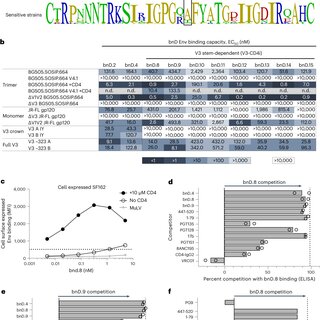
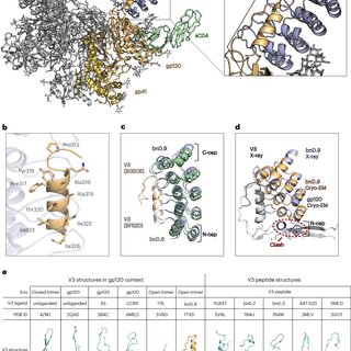
Trapping the HIV-1 V3 loop in a helical conformation enables broad neutralization

August 2023

·

85 Reads

·

1 Citation

Nature Structural & Molecular Biology

The third variable (V3) loop on the human immunodeficiency virus 1 (HIV-1) envelope glycoprotein trimer is indispensable for virus cell entry. Conformational masking of V3 within the trimer allows efficient neutralization via V3 only by rare, broadly neutralizing glycan-dependent antibodies targeting the closed prefusion trimer but not by abundant antibodies that access the V3 crown on open trimers after CD4 attachment. Here, we report on a distinct category of V3-specific inhibitors based on designed ankyrin repeat protein (DARPin) technology that reinstitute the CD4-bound state as a key neutralization target with up to >90% breadth. Broadly neutralizing DARPins (bnDs) bound V3 solely on open envelope and recognized a four-turn amphipathic α-helix in the carboxy-terminal half of V3 (amino acids 314–324), which we termed ‘αV3C’. The bnD contact surface on αV3C was as conserved as the CD4 binding site. Molecular dynamics and escape mutation analyses underscored the functional relevance of αV3C, highlighting the potential of αV3C-based inhibitors and, more generally, of postattachment inhibition of HIV-1.


HIV-1 neutralizing antibodies elicited in humans by a prefusion-stabilized envelope trimer form a reproducible class targeting fusion peptide

July 2023

·

60 Reads

·

5 Citations

Cell Reports

Elicitation of antibodies that neutralize the tier-2 neutralization-resistant isolates that typify HIV-1 transmission has been a long-sought goal. Success with prefusion-stabilized envelope trimers eliciting autologous neutralizing antibodies has been reported in multiple vaccine-test species, though not in humans. To investigate elicitation of HIV-1 neutralizing antibodies in humans, here, we analyze B cells from a phase I clinical trial of the "DS-SOSIP"-stabilized envelope trimer from strain BG505, identifying two antibodies, N751-2C06.01 and N751-2C09.01 (named for donor-lineage.clone), that neutralize the autologous tier-2 strain, BG505. Though derived from distinct lineages, these antibodies form a reproducible antibody class that targets the HIV-1 fusion peptide. Both antibodies are highly strain specific, which we attribute to their partial recognition of a BG505-specific glycan hole and to their binding requirements for a few BG505-specific residues. Prefusion-stabilized envelope trimers can thus elicit autologous tier-2 neutralizing antibodies in humans, with initially identified neutralizing antibodies recognizing the fusion-peptide site of vulnerability.


Push-pull mechanics of E-cadherin ectodomains in biomimetic adhesions

July 2023

·

13 Reads

·

1 Citation

Biophysical Journal

E-cadherin plays a central role in cell-cell adhesion. The ectodomains of wild type cadherins form a crystalline-like two dimensional lattice in cell-cell interfaces mediated by both trans (apposed cell) and cis (same cell) interactions. In addition to these extracellular forces, adhesive strength is further regulated by cytosolic phenomena involving α and β-catenin-mediated interactions between cadherin and the actin cytoskeleton. Cell-cell adhesion can be further strengthened under tension through mechanisms that have not been definitively characterized in molecular detail. Here we quantitatively determine the role of the cadherin ectodomain in mechanosensing. To this end, we devise an E-cadherin-coated emulsion system, in which droplet surface tension is balanced by protein binding strength to give rise to stable areas of adhesion. To reach the honeycomb/cohesive limit, an initial emulsion compression by centrifugation facilitates E-cadherin trans-binding, while a high protein surface concentration enables the cis-enhanced stabilization of the interface. We observe an abrupt concentration dependence on recruitment into adhesions of constant crystalline density, reminiscent of a first-order phase transition. Removing the lateral cis-interaction with a "cis mutant" shifts this transition to higher surface densities leading to denser, yet weaker adhesions. In both proteins, the stabilization of progressively larger areas of deformation is consistent with single-molecule experiments that show a force-dependent lifetime enhancement in the cadherin ectodomain, which may be attributed to the "X-dimer" bond.


Citations (78)


... in close proximity to the receptor binding motif [9][10][11] , blocking the interaction of RBD with the cellular SARS-CoV-2 receptor, angiotensinconverting enzyme 2 (ACE2) 12 , although a few bind elsewhere and may function to destabilize the S trimer [13][14][15] . A number of potent mAbs bind to a so-called supersite in the N-terminal domain (NTD) of S1, although their mechanism of neutralization is poorly understood 16,17 and some bind at the interface of the NTD and SD1 locking the RBDs down to prevent ACE2 interaction 18 . ...

Reference:

The SARS-CoV-2 neutralizing antibody response to SD1 and its evasion by BA.2.86
Antibodies targeting a quaternary site on SARS-CoV-2 spike glycoprotein prevent viral receptor engagement by conformational locking
  • Citing Article
  • September 2023

Immunity

... This was shown in the case of the DS-2 molecule described by ref. 42, where the successful incorporation of an additional disulfide bond to DS-Cav1 achieved 4X greater neutralizing antibody titers. Similarly, Ou et al. reported a triple disulfide-bonded metapneumovirus F protein that achieved 10-fold higher titers than the single disulfide-bonded version of the protein 59 . There may, however, be a virus or protein-specific upper limit to potency improvements as incorporation of an additional disulfide bond in an otherwise single-disulfide bond stabilized parainfluenza virus type 3 fusion protein did not further improve neutralization titers 60,61 . ...

Structure-based design of a single-chain triple-disulfide-stabilized fusion-glycoprotein trimer that elicits high-titer neutralizing responses against human metapneumovirus

... Owing to their design, standard DARPins lack the option of gaining access to constrained regions by elongating loops as the synthetic library used here does not encode them 44 , even though such constructs have been made 59 . Typically, the synthetic DARPin libraries will not contain short insertions and deletions nor loss of individual repeats, but occasionally such clones derived by PCR errors are amplified when no other solution to high-affinity binding may exist, e.g., by steric restriction 60,61 . ...

Trapping the HIV-1 V3 loop in a helical conformation enables broad neutralization

Nature Structural & Molecular Biology

... To con rm the epitope and explore the binding mode of bNAb4251, we decided to perform cryo-electron microscopy (cryo-EM) of the antigen-binding fragment (Fab)4251 in complex with the soluble native-like trimer (BG505 DS-SOSIP) 51 . After several rounds of 2D and 3D classi cations (Extended Data Fig 9), we could segregate trimers with zero, or one Fab attached and solved the structure of the complex at the resolution of 3.7Å (Figure 5a and, Supplementary Table 3). ...

HIV-1 neutralizing antibodies elicited in humans by a prefusion-stabilized envelope trimer form a reproducible class targeting fusion peptide

Cell Reports

... FEP calculations are shown to be highly effective in predicting the ligand binding affinities in many systems [56][57][58][59]. For example, FEP has been applied to study the effect of mutation on protein-protein interactions particularly in the context of SARS-CoV-2 RBD: ACE2 binding affinity [60], identify novel allosteric inhibitor of human transcription factor [61] and discover Nirmatrelvir resistance mutations in SARS-CoV2 3CLpro [62]. Other variants of FEP have started to yield significant improvements in computing accurate relative binding free energy of congeneric ligands. ...

Free Energy Perturbation Calculations of Mutation Effects on SARS-CoV-2 RBD::ACE2 Binding Affinity
  • Citing Article
  • June 2023

Journal of Molecular Biology

... Conversely, RBD-specific antibodies of classes 3 and 4 are non-ACE2 blocking antibodies that bind 'up' and 'down' (class 3) or only 'up' (class 4). Multiple factors contribute to an enhanced frequency of mutations in the RBD, thereby influencing vaccine immunogenicity and monoclonal antibody recognition [8]. The N-terminal domain (NTD) upstream of the RBD in the S1 subunit, similarly known to elicit potent neutralizing antibodies [9][10][11], is also a region of highly mutated residues primarily centered around a supersite located in the N3 loop (residues 141-156) [9,11]. ...

Antibodies that neutralize all current SARS-CoV-2 variants of concern by conformational locking

... The crystal structures of megalin (PDB ID: 8EM4) were obtained from the protein data bank database (PDB) (Beenken et al. 2023). The high ambiguity-driven protein-protein docking (HADDOCK) v2.4 platform was used for computational biomolecular interaction studies (protein-peptide). ...

Structures of LRP2 reveal a molecular machine for endocytosis

... In recent years, several studies have revealed that broadly neutralizing antibodies against HIV-1 are a class of preventive antiviral drugs with advantages of high efficacy and few side effects, and they have shown promising antiviral effects in laboratory and clinical studies [26][27][28][29], indicating that these may be used as a supplement to the current antiretroviral HIV-1 drugs. A large number of HIV-1 bNAbs have been developed, including bNAbs that recognize various conserved epitopes on HIV-1 envelope glycoprotein, bNAbs that recognize HIV-1 cell receptors and co-receptors, as well as bispecific and trispecific antibodies engineered based on the above single antibodies [30][31][32]. One of the main reasons why HIV-1 can pose a significant challenge to humans is that HIV-1 is very easy to mutate and has many subtypes. ...

Bispecific antibody CAP256.J3LS targets V2-apex and CD4-binding sites with high breadth and potency

... These results suggest that GAPDH (a host protein) could harbor the potential for use as a therapeutic agent to inhibit the viral entry for up to 12 h post-application. In future, this could possibly provide leads for the further development of peptide/protein-based blockers of viral infections, which could be deployed by way of upper-respiratory tracts sprays [65,66]. In silico molecular docking analysis indicated that primarily basic amino acid residues of GAPDH interact with different amino acids in the B chain of the viral S protein. ...

A potent and broad neutralization of SARS-CoV-2 variants of concern by DARPins

Nature Chemical Biology

... The initial atomistic structures of the "one RBD up" (PDB ID: 6XM3) [28] and "RBD down" (PDB ID: 6XR8) [29] conformations were downloaded from the Protein Data Bank (PDB). These two high-resolution structures contained most of the S protein architecture, yet there are still some residues missing. ...

Cryo-EM Structures Delineate a pH-Dependent Switch that Mediates Endosomal Positioning of SARS-CoV-2 Spike Receptor-Binding Domains
  • Citing Article
  • January 2020

SSRN Electronic Journal