Heidi Koegel's research while affiliated with Eawag: Das Wasserforschungs-Institut des ETH-Bereichs and other places

What is this page?


This page lists the scientific contributions of an author, who either does not have a ResearchGate profile, or has not yet added these contributions to their profile.

It was automatically created by ResearchGate to create a record of this author's body of work. We create such pages to advance our goal of creating and maintaining the most comprehensive scientific repository possible. In doing so, we process publicly available (personal) data relating to the author as a member of the scientific community.

If you're a ResearchGate member, you can follow this page to keep up with this author's work.

If you are this author, and you don't want us to display this page anymore, please let us know.

Publications (11)


Figure S3
  • Data
  • File available

May 2009

·

7 Reads

Aleksandra Piwko-Czuchra

·

Heidi Koegel

·

·

[...]

·

Reinhard Fässler

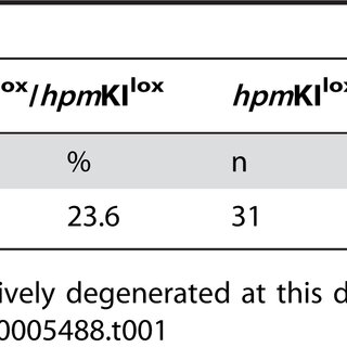
Wound healing in hpm mice. (A) Morphometrical analysis of wound healing parameters of 5 day wounds. Slightly reduced wound closure in hpm mice (control: n = 19, N = 7; hpm: n = 21, N = 7). The area of the hyperproliferative epithelium (HE) was similar in 5 day wounds of control and hpm mice (control: n = 14, N = 6; hpm: n = 11, N = 6). (B) Morphometrical analysis of wound healing parameters 13 days after wounding. The wound width was significantly increased (p = 0.0005; control: n = 16, N = 5; hpm: n = 16, N = 5) and the epidermis still hyperthickened in 13 day wounds of hpm mice (p = 0.0013; control: n = 19, N = 7; hpm: n = 21, N = 7). (C) 13 day wounds were examined for lacZ expression. Black arrowheads indicate the edges of the wound, white arrowheads mark an area of lacZ negative keratinocytes in the middle of the wound epithelium (control: N = 3; hpm: N = 4). Boxed area is shown at higher magnification. n, number of measurements; N, number of mice. (6.50 MB TIF)

Download
Share

Figure S1

May 2009

·

8 Reads

Targeting strategy for the hpm allele. (A) Partial map of the β1A wild-type (wt), the hpm knock-in allele before (hpmKIneo-tk+) and after Cre-mediated deletion of the floxed neo-tk cassette (hpmKIlox). External probes used for Southern blotting after BamHI digest and genotyping PCR primers are indicated. Cyto-cDNA, a wt 141 bp cDNA fragment coding for the complete cytoplasmic domain of the β1A integrin gene including the endogenous stop codon; E15cyto, endogenous sequence of exon 15 encoding for the membrane proximal part of the β1 integrin cytoplasmic tail; filled boxes, exons; D, exon D; neo, neomycin resistance gene; tk, thymidine kinase gene; triangles, loxP sites; pA, polyadenylation signal. (B) Left panel: Southern blot analysis with the 5′external probe demonstrating recombination of the targeting construct (hpmneo-tk) into the wt β1 integrin locus giving rise to a 22.9 kb wt band and a 7 kb recombinant band. Right panel: Southern blot analysis with the 3′external probe demonstrating the deletion of the neo-tk cassette from the knockin hpmKIneo-tk+ allele (hpmKIlox) with a 22.9 kb wt band and 16 kb hpmKIlox band. (C) Representative PCR on genomic DNA from tail snips of offspring from hpmKIlox/+ intercrosses using a forward primer (primer S) binding to the transmembrane coding sequence of the β1 integrin gene, within exon 15 and a reverse primer (primer AS) binding to an intronic sequence downstream of exon 15. The 423 and 191 bp DNA bands correspond to the hpmKIlox and the wt β1 integrin allele, respectively. (0.95 MB TIF)


Figure S2

May 2009

·

8 Reads

Integrin profile of hpm keratinocytes. Cell surface expression of integrins in freshly isolated keratinocytes from control and hpm mice at indicated ages were assessed by flow cytometry. Keratinocytes were stained with antibodies against α2, β4 and α6 integrins. Expression of integrin subunits was normalized to the expression level of age-matched controls. Cell surface expression of α2 integrin is significantly reduced in 3-week-old hpm mice and increases to control levels in 20-month-old hpm mice. Expression of α6 and β4 integrin in young and old hpm mutant mice is increased compared to controls (middle and lower panel, respectively). Mean fluorescence intensities were corrected for background fluorescence. Error bars indicate s.d. At least 2 control and 3 hpm mutant mice per developmental stage were analysed. (0.90 MB TIF)


Figure 2 Spontaneous recombination of the floxed Srf allele in Srf flex1/flex1 K14CrePR1 mice. (A) Schematic illustration of Srf alleles and genotyping of Srf flex1/flex1 × Srf WT/flex1 K14CrePR1 offspring: Srf flex1/flex1 K14CrePR1 (Srf mutant) mice were identified by a 730-bp fragment (Srf flex1 ), a 412-bp fragment (Cre recombinase), and the absence of the 650-bp fragment that identifies the WT allele (Srf WT ). A 380-bp fragment was amplified from the recombined Srf allele (Srf lx ). E/R and L/R indicate primer pairs described in ref. 15. (B) A representative Western blot of total back skin lysates showing reduced SRF protein levels in the skin of Srf flex1/flex1 K14CrePR1 mice at P14 compared with skin of control littermates. GAPDH was used as a loading control. (C–E) Macroscopic appearance of control and Srf mutant mice at P14. Note the scaly lesions on the back (C), the paws (D), and the tail (E). (F) Immunohistochemical analysis of SRF in back skin from Srf mutant and control mice at P2 shows comparable SRF expression patterns. (G) At P14, nuclear SRF staining is markedly reduced in the epidermis of Srf flex1/flex1 K14CrePR1 mice. (F and G) Dotted lines indicate the basement membrane. Scale bars: 50 μm. Sections were counterstained with hematoxylin.  
Figure 3  
Figure 4 Disruption of the actin cytoskeleton and reduced cell-cell and cell-matrix contacts in Srf mutant mice. (A) Phalloidin-FITC staining of skin sections of control and Srf mutant mice at P14 shows markedly reduced levels of polymerized actin (F-actin) in lesions of Srf mutant skin (upper panels). Immunofluorescence staining for E-cadherin shows membrane localization in skin of control and Srf mutant mice (lower panels). Dotted lines indicate the basement membrane. Scale bar: 20 μm. (B–E) Ultrastructural analysis of control and Srf mutant epidermis at P14. (B) Keratinocytes in the basal cell layer of lesional Srf mutant epidermis are strongly enlarged and lack most cell-cell contacts compared with control skin, in which basal keratinocytes are small and densely packed. Arrows indicate the basement membrane. (C) Stratum corneum (sc) of lesional Srf mutant epidermis is thicker and less compact than stratum corneum of control epidermis. (D) Epidermal lesions of Srf mutant skin show areas with defective hemidesmosomes (arrows). Note that collagen bundles below the basement membrane are absent in Srf mutant skin. (E) Desmosomes (arrows) are markedly reduced in Srf mutant skin, and cell-cell contacts are largely absent. (F and G) Morphometric analysis confirmed a distinct reduction of the number of desmosomes (F), while no change of the mean length of single desmosomes could be found (G). Error bars indicate SD. Scale bars: 5 μm (B and C); 1 μm (D and E). ***P ≤ 0.001.  
Figure 5 Keratinocyte differentiation is impaired in Srf mutant epidermis. (A and B) Immunohistochemical analysis of tail-skin sections from control and Srf mutant mice (P11–P15) for the expression of epidermal differentiation markers. (A) Expression of K14, K15, and K5 was reduced and their localization was irregular in Srf mutant tail skin. Expression of the differentiation markers K10 and loricrin was delayed and reduced compared with that in control skin. (B) Srf mutant mice show abnormal interfollicular expression of K6, K17, and K8/K18. Sections were counterstained with hematoxylin. Scale bar: 100 μm.  
Figure 6 Skin lesions of Srf mutant mice reveal markers of inflammatory skin disease. (A) Immunofluorescence analysis of skin sections shows high suprabasal expression of the β1 integrin (upper panels) and α-integrin (lower panels) subunits in Srf mutant skin. Scale bars: 20 μm. Dotted lines indicate the basement membrane. (B) p-STAT3 levels are markedly increased in total skin lysates from Srf mutant mice. GAPDH was used as a loading control. (C) Relative mRNA levels of Srf, IL-1β, S100A8, and S100A9 in 5 individual Srf mutant mice are shown (1–3, P14; 4, P13: severe scaling phenotype; 5, P42: slowly progressing phenotype). Reduced Srf expression correlates with increased IL-1β, S100A8 and S100A9 mRNA levels. RNA levels of a control littermate are set as 1. mRNA of the housekeeping gene Gapdh was used for normalization. PCR analysis was performed in duplicate, and the average of both values is shown. Each result was reproduced in an independent experiment.  

+1

Loss of serum response factor in keratinocytes results in hyperproliferative skin disease in mice

May 2009

·

421 Reads

·

61 Citations

The Journal of clinical investigation

The transcription factor serum response factor (SRF) plays a crucial role in the development of several organs. However, its role in the skin has not been explored. Here, we show that keratinocytes in normal human and mouse skin expressed high levels of SRF but that SRF expression was strongly downregulated in the hyperproliferative epidermis of wounded and psoriatic skin. Keratinocyte-specific deletion within the mouse SRF locus during embryonic development caused edema and skin blistering, and all animals died in utero. Postnatal loss of mouse SRF in keratinocytes resulted in the development of psoriasis-like skin lesions. These lesions were characterized by inflammation, hyperproliferation, and abnormal differentiation of keratinocytes as well as by disruption of the actin cytoskeleton. Ultrastructural analysis revealed markedly reduced cell-cell and cell-matrix contacts and loss of cell compaction in all epidermal layers. siRNA-mediated knockdown of SRF in primary human keratinocytes revealed that the cytoskeletal abnormalities and adhesion defects were a direct consequence of the loss of SRF. In contrast, the hyperproliferation observed in vivo was an indirect effect that was most likely a consequence of the inflammation. These results reveal that loss of SRF disrupts epidermal homeostasis and strongly suggest its involvement in the pathogenesis of hyperproliferative skin diseases, including psoriasis.


Keratinocyte-derived follistatin regulates epidermal homeostasis and wound repair

February 2009

·

478 Reads

·

26 Citations

Laboratory Investigation

Activin is a growth and differentiation factor that controls development and repair of several tissues and organs. Transgenic mice overexpressing activin in the skin were characterized by strongly enhanced wound healing, but also by excessive scarring. In this study, we explored the consequences of targeted activation of activin in the epidermis and hair follicles by generation of mice lacking the activin antagonist follistatin in keratinocytes. We observed enhanced keratinocyte proliferation in the tail epidermis of these animals. After skin injury, an earlier onset of keratinocyte hyperproliferation at the wound edge was observed in the mutant mice, resulting in an enlarged hyperproliferative epithelium. However, granulation tissue formation and scarring were not affected. These results demonstrate that selective activation of activin in the epidermis enhances reepithelialization without affecting the quality of the healed wound.


β1 Integrin-Mediated Adhesion Signalling Is Essential for Epidermal Progenitor Cell Expansion

February 2009

·

325 Reads

·

49 Citations

PLOS ONEPLOS ONE

There is a major discrepancy between the in vitro and in vivo results regarding the role of beta1 integrins in the maintenance of epidermal stem/progenitor cells. Studies of mice with skin-specific ablation of beta1 integrins suggested that epidermis can form and be maintained in their absence, while in vitro data have shown a fundamental role for these adhesion receptors in stem/progenitor cell expansion and differentiation. To elucidate this discrepancy we generated hypomorphic mice expressing reduced beta1 integrin levels on keratinocytes that developed similar, but less severe defects than mice with beta1-deficient keratinocytes. Surprisingly we found that upon aging these abnormalities attenuated due to a rapid expansion of cells, which escaped or compensated for the down-regulation of beta1 integrin expression. A similar phenomenon was observed in aged mice with a complete, skin-specific ablation of the beta1 integrin gene, where cells that escaped Cre-mediated recombination repopulated the mutant skin in a very short time period. The expansion of beta1 integrin expressing keratinocytes was even further accelerated in situations of increased keratinocyte proliferation such as wound healing. These data demonstrate that expression of beta1 integrins is critically important for the expansion of epidermal progenitor cells to maintain epidermal homeostasis.


Overexpression of mIGF-1 in Keratinocytes Improves Wound Healing and Accelerates Hair Follicle Formation and Cycling in Mice

November 2008

·

83 Reads

·

59 Citations

American Journal Of Pathology

Insulin-like growth factor 1 (IGF-1) is an important regulator of growth, survival, and differentiation in many tissues. It is produced in several isoforms that differ in their N-terminal signal peptide and C-terminal extension peptide. The locally acting isoform of IGF-1 (mIGF-1) was previously shown to enhance the regeneration of both muscle and heart. In this study, we tested the therapeutic potential of mIGF-1 in the skin by generating a transgenic mouse model in which mIGF-1 expression is driven by the keratin 14 promoter. IGF-1 levels were unchanged in the sera of hemizygous K14/mIGF-1 transgenic animals whose growth was unaffected. A skin analysis of young animals revealed normal architecture and thickness as well as proper expression of differentiation and proliferation markers. No malignant tumors were formed. Normal homeostasis of the putative stem cell compartment was also maintained. Healing of full-thickness excisional wounds was accelerated because of increased proliferation and migration of keratinocytes, whereas inflammation, granulation tissue formation, and scarring were not obviously affected. In addition, mIGF-1 promoted late hair follicle morphogenesis and cycling. To our knowledge, this is the first work to characterize the simultaneous, stimulatory effect of IGF-1 delivery to keratinocytes on two types of regeneration processes within a single mouse model. Our analysis supports the use of mIGF-1 for skin and hair regeneration and describes a potential cell type-restricted action.


Cell cycle inhibitors p21 and p16 are required for the regulation of Schwann cell proliferation

January 2006

·

147 Reads

·

43 Citations

Glia

Regulated cell proliferation is a crucial prerequisite for Schwann cells to achieve myelination in development and regeneration. In the present study, we have investigated the function of the cell cycle inhibitors p21 and p16 as potential regulators of Schwann cell proliferation, using p21- or p16-deficient mice. We report that both inhibitors are required for proper withdrawal of Schwann cells from the cell cycle during development and following injury. Postnatal Schwann cells express p21 exclusively in the cytoplasm, first detectable at postnatal day 7. This cytoplasmic p21 expression is necessary for proper Schwann cell proliferation control in the late development of peripheral nerves. After axonal damage, p21 is found in Schwann cell nuclei during the initiation of the proliferation period. This stage is critically regulated by p21, since loss of p21 leads to a strong increase in Schwann cell proliferation. Unexpectedly, p21 levels are upregulated in this phase suggesting that the role of p21 may be more complex than purely inhibitory for the Schwann cell cycle. However, inhibition of Schwann cell proliferation is the overriding crucial function of p21 and p16 in peripheral nerves as revealed by the consequences of loss-of-function in development and after injury. Different mechanisms appear to underlie the inhibitory function, depending on whether p21 is cytoplasmic or nuclear.


Confocal ratiometric voltage imaging of cultured human keratinocytes reveals layer-specific responses to ATP

May 2003

·

24 Reads

·

14 Citations

AJP Cell Physiology

Recent evidence suggests that changes in membrane potential influence the proliferation and differentiation of keratinocytes. To further elucidate the role of changes in membrane potential for their biological fate, the electrical behavior of keratinocytes needs to be studied under complex conditions such as multilayered cultures. However, electrophysiological recordings from cells in the various layers of a complex culture would be extremely difficult. Given the high spatial resolution of confocal imaging and the availability of novel voltage-sensitive dyes, we combined these methods in an attempt to develop a viable alternative for recording membrane potentials in more complex tissue systems. As a first step, we used confocal ratiometric imaging of fluorescence resonance energy transfer (FRET)-based voltage-sensitive dyes. We then validated this approach by comparing the optically recorded voltage signals in HaCaT keratinocytes with the electrophysiological signals obtained by whole cell recordings of the same preparation. We demonstrate 1) that optical recordings allow precise multisite measurements of voltage changes evoked by the extracellular signaling molecules ATP and bradykinin and 2) that responsiveness to ATP differs in various layers of cultured keratinocytes.


Unexpected down-regulation of the hIK1 Ca2+-activated K+ channel by its opener 1-ethyl-2-benzimidazolinone in HaCaT Keratinocytes - Inverse effects on cell growth and proliferation

February 2003

·

30 Reads

·

39 Citations

Journal of Biological Chemistry

We used a combination of electrophysiological and cell and molecular biological techniques to study the regulation and functional role of the intermediate conductance Ca2+-activated K+ channel, hIK1, in HaCaT keratinocytes. When we incubated cells with the hIK1 opener, 1-ethyl-2-benzimidazolinone (1-EBIO), to investigate the cellular consequences of prolonged channel activity, an unexpected down-regulation of channels occurred within a few hours. The same effect was produced by the hIK1 openers chlorzoxazone and zoxazolamine and was also observed in a different cell line (C6 glioma cells). After 3 days of treatment with 1-EBIO, mRNA levels of hIK1 were substantially diminished and no channel activity was detected. Down-regulation of hIK1 was accompanied by a loss of mitogenic activity and a strong increase in cell size. After withdrawal of 1-EBIO, hIK1 mRNA and channel activity fully recovered and the cells resumed mitogenic activity. Our data present evidence for a novel feedback mechanism of hIK1 expression that appears to result from the paradoxical action of its pharmacological activator during prolonged application. Because the down-regulation of hIK1 bears immediate significance on the biological fate of keratinocytes, 1-EBIO and related compounds might emerge as potent tools to influence the proliferation of various non-excitable cells endowed with IK channels.


Citations (8)


... In this study, there is a limitation in addressing whether the reduction level of ITGB1 in EpiSCs with DSBs ( Figures S4F and S4G) is sufficient for their delamination. As there is evidence that ITGB1 low EpiSCs are replaced by ITGB1 high EpiSCs in vivo (Piwko-Czuchra et al., 2009), our data suggest the involvement of ITGB1 in the stem cell selection mechanism to safeguard the epithelial genomic quality. ...

Reference:

Dynamic stem cell selection safeguards the genomic integrity of the epidermis
β1 Integrin-Mediated Adhesion Signalling Is Essential for Epidermal Progenitor Cell Expansion

... Further, siRNA-mediated knockdown of SRF in primary 266 human keratinocytes revealed that cytoskeletal abnormalities and adhesion defects were direct consequences of SRF loss. 81 Inhibition of Polycystin 1 (PC1) function was associated with increased cell proliferation and migration in human keratinocytes. 82 Furthermore, overexpression of Granulin Precursor (PGRN) has been shown to inhibit keratinocyte inflammation by negatively regulating the production of inflammatory factors and positively influencing autophagy. ...

Loss of serum response factor in keratinocytes results in hyperproliferative skin disease in mice

The Journal of clinical investigation

... Furthermore, epidermal thickness and procollagen type 1 c-peptide production were increased in 3D skin equivalents from old donors, thereby shifting an old skin phenotype towards a juvenile phenotype. In line with these results, mice lacking the Activin antagonist Fst have been shown to exhibit enhanced keratinocyte proliferation (Antsiferova et al., 2009). Furthermore, collagen synthesis is stimulated in mice overexpressing Activin A in keratinocytes (Wietecha et al., 2020). ...

Keratinocyte-derived follistatin regulates epidermal homeostasis and wound repair

Laboratory Investigation

... 35 The effect of reepithelialization at the wound site and proliferation in hair follicles has been demonstrated. 36 Insulin-like growth factor plays a role as a progressive factor in the regeneration of fibroblast-derived tissues. Insulin-like growth factor can trigger angiogenesis via VEGF. ...

Overexpression of mIGF-1 in Keratinocytes Improves Wound Healing and Accelerates Hair Follicle Formation and Cycling in Mice
  • Citing Article
  • November 2008

American Journal Of Pathology

... Various types of Ca 2+ -activated K + (K Ca ) channels are known to be expressed in human keratinocytes. These include BK Ca channels in oral keratinocytes and HaCaT cells [16,17], the intermediate conductance Ca 2+ -activated K + channel (hIK1) in HaCaT cells and epidermal keratinocytes [14,18], and the small conductance Ca 2+ -activated K + channel type 4 (SK4) in HaCaT cells [19]. However, there are few reports on the expression of BK Ca channels in NHEKs rather than cell lines. ...

Expression and biological significance of Ca2+-activated ion channels in human keratinocytes
  • Citing Article
  • February 2001

The FASEB Journal

... Conversely, K + channel inhibition has been found to accelerate wound healing in intestinal epithelial cells [9,11]. Keratinocytes express various K + channels, including ATP-sensitive, two-pore domain, and Ca 2+ -independent/dependent K + channels [12][13][14][15]. However, the modulation and function of these channels in normal human epidermal keratinocytes (NHEKs) remain underexplored. ...

Unexpected down-regulation of the hIK1 Ca2+-activated K+ channel by its opener 1-ethyl-2-benzimidazolinone in HaCaT Keratinocytes - Inverse effects on cell growth and proliferation

Journal of Biological Chemistry

... Time difference between the right and left eye CC2-DMPE signals was measured by taking images at regular intervals of 10 minutes. While CC2-DMPE used alone cannot precisely quantify V mem levels, the strong fluorescence from this positively charged dye has been shown previously to reliably identify hyperpolarized regions as confirmed by electrophysiological impalement [65][66][67]136] and to identify the same locations of hyperpolarized cells in the early face as did prior studies using ratiometric imaging [69]. ...

Confocal ratiometric voltage imaging of cultured human keratinocytes reveals layer-specific responses to ATP
  • Citing Article
  • May 2003

AJP Cell Physiology

... Mutations in EGR2 have been identified to lead to inherited peripheral neuropathies, including Charcot-Marie-Tooth Type 1 (Šafka Brožková et al., 2012), a demyelinating form associated with dysregulated Schwann cell proliferation and cell cycle exit (Atanasoski et al., 2006). Accumulating evidence indicates that EGR2, a transcription factor, plays the role of regulator in these processes (Decker, 2006;Topilko et al., 1994;Zorick et al., 1996) and has been shown to directly bind to the p21 promoter in myelinating rat sciatic nerve (Srinivasan et al., 2012). ...

Cell cycle inhibitors p21 and p16 are required for the regulation of Schwann cell proliferation
  • Citing Article
  • January 2006

Glia