Berge A. Minassian's research while affiliated with University of Texas Southwestern Medical Center and other places

What is this page?


This page lists the scientific contributions of an author, who either does not have a ResearchGate profile, or has not yet added these contributions to their profile.

It was automatically created by ResearchGate to create a record of this author's body of work. We create such pages to advance our goal of creating and maintaining the most comprehensive scientific repository possible. In doing so, we process publicly available (personal) data relating to the author as a member of the scientific community.

If you're a ResearchGate member, you can follow this page to keep up with this author's work.

If you are this author, and you don't want us to display this page anymore, please let us know.

Publications (346)


Embryonic lethality of Gde5−/− mice and higher GPC level in skeletal muscle and liver of Gde5+/− mice
a PCR genotyping using DNA from single blastocyst. Isolated WT, Gde5+/− and Gde5−/− mutant blastocysts. b–d Choline metabolites levels in skeletal muscle of Gde5+/− and WT mice fed with standard AIN93 diet. e–g Choline metabolites levels in livers of Gde5+/− and WT mice fed with standard AIN93 diet. hGde5 mRNA expression on various tissues in WT mice. Values are means ± SEM. Statistical analysis was performed with Student’s t test. *p < 0.05.
Abnormal GPC/choline metabolism and reduced passive force in the skeletal muscle of Gde5 skKO mice
a Western blot, mRNA expression, and GPC hydrolyzing activity in skeletal muscle of Gde5 skKO and WT mice. b–f Choline metabolites levels in skeletal muscle (b), liver (c), kidney (d), white adipose tissue (e), and serum (f) of Gde5 skKO and WT mice. g Schematic figure of in vivo model for contractile force and fatigability experiment. h In vivo contractile force test of Gde5 skKO and WT mice (n = 7). i Representative examples of contractile force test. j Fatigability test of Gde5 skKO and WT mice (n = 7). Values are means ± SEM. Statistical analysis was performed with Student’s t test (a–f) and two-way ANOVA (h, j). *p < 0.05; **p < 0.01; ***p < 0.001; ****p < 0.0001; #p < 0.05 (main effect).
Abnormal glucose metabolism in the skeletal muscle of Gde5 skKO mice
a Volcano plot of water-soluble metabolites in Gde5 skKO and WT skeletal muscle. b Heatmap representation of glucose metabolism-related metabolites levels in Gde5 skKO and WT skeletal muscle. c Glucose metabolism-related metabolites levels in Gde5 skKO and WT skeletal muscle (n = 6–7). d Amino acids and their related metabolites levels in Gde5 skKO and WT skeletal muscle (n = 6–7). e Venn diagram of DEGs between Gde5 skKO under fasting and 1 h-post 20% glucose injection. f Hexokinase activity of Gde5 skKO and WT skeletal muscle. g Insulin tolerance test of Gde5 skKO and WT mice under normal chow diet (n = 7). h Fasting blood glucose levels of Gde5 skKO and WT mice (n = 7) under chow diet and high-fat diet. ND, normal chow diet; HFD, high-fat diet. i Insulin tolerance test of Gde5 skKO and WT mice under high-fat diet (n = 7). j Glycogen level of Gde5 skKO and WT skeletal muscle. Values are means ± SEM. Statistical analysis was performed with Student’s t test (c, d, f, h, j) and two-way ANOVA (g, i). #p < 0.05 (main effect); *p < 0.05; **p < 0.01; ***p < 0.001; ****p < 0.0001.
Comparison of phospholipid profile of the skeletal muscle from Gde5 skKO mice with denervated and mdx mice
a Volcano plot of phospholipid content in Gde5 skKO and WT mice skeletal muscle. b–d Profiles of PC (b), PE (c), and LPC (d) molecular species in Gde5 skKO and WT mice skeletal muscle (n = 7). e Heatmap representation of phospholipid species in skeletal muscle of Gde5 skKO, mdx, denervation and their respective controls. f, g Profiles of PC molecular species in denervated (f) and mdx (g) skeletal muscle. h TAG molecular species of Gde5 skKO and WT skeletal muscle under normal chow diet. TAG, triacylglycerol. Values are means ± SEM. Statistical analysis was performed with Student’s t test. *p < 0.05; **p < 0.01; ***p < 0.001; ****p < 0.0001.
Role of GPC in modulating PC composition of the skeletal muscle
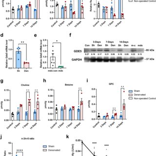
a–c PC compositional change post-denervation with non-operated control as external control and sham-operated muscle as internal control. d, e qPCR analysis of Gde5 mRNA expression in denervated (d) and mdx (e) skeletal muscle compared to their respective control. Sh, sham; Den, denervated; mdx-con, mdx control. f GDE5 western blot on denervated and mdx skeletal muscle compared to their respective control. Con, non-operated control; Sh, sham; Den, denervated; m-c, mdx control. g–i Choline metabolites levels post-denervation with non-operated control as external control and sham-operated muscle as internal control. j n-3/n-6 ratio of denervated Gde5 skKO and WT skeletal muscle compared to respective sham-operated muscle. k PLA2 activity in skeletal muscle homogenates following addition of GPC (n = 6). Values are means ± SEM. Statistical analysis was performed with Student’s t test. *p < 0.05; **p < 0.01; ***p < 0.001; ****p < 0.0001.

+3

GDE5/Gpcpd1 activity determines phosphatidylcholine composition in skeletal muscle and regulates contractile force in mice
  • Article
  • Full-text available

May 2024

·

21 Reads

Communications Biology

Rahmawati Aisyah

·

Noriyasu Ohshima

·

Daiki Watanabe

·

[...]

·

Glycerophosphocholine (GPC) is an important precursor for intracellular choline supply in phosphatidylcholine (PC) metabolism. GDE5/Gpcpd1 hydrolyzes GPC into choline and glycerol 3-phosphate; this study aimed to elucidate its physiological function in vivo. Heterozygous whole-body GDE5-deficient mice reveal a significant GPC accumulation across tissues, while homozygous whole-body knockout results in embryonic lethality. Skeletal muscle-specific GDE5 deletion (Gde5 skKO) exhibits reduced passive force and improved fatigue resistance in electrically stimulated gastrocnemius muscles in vivo. GDE5 deficiency also results in higher glycolytic metabolites and glycogen levels, and glycerophospholipids alteration, including reduced levels of phospholipids that bind polyunsaturated fatty acids (PUFAs), such as DHA. Interestingly, this PC fatty acid compositional change is similar to that observed in skeletal muscles of denervated and Duchenne muscular dystrophy mouse models. These are accompanied by decrease of GDE5 expression, suggesting a regulatory role of GDE5 activity for glycerophospholipid profiles. Furthermore, a DHA-rich diet enhances contractile force and lowers fatigue resistance, suggesting a functional relationship between PC fatty acid composition and muscle function. Finally, skinned fiber experiments show that GDE5 loss increases the probability of the ryanodine receptor opening and lowers the maximum Ca²⁺-activated force. Collectively, GDE5 activity plays roles in PC and glucose/glycogen metabolism in skeletal muscle.

Download
Share


1H and 31P magnetic resonance spectroscopy reveal potential pathogenic and biomarker metabolite alterations in Lafora disease

March 2024

·

8 Reads

Brain Communications

Lafora disease is a fatal teenage-onset progressive myoclonus-epilepsy and neurodegenerative disease associated with polyglucosan bodies. Polyglucosans are long-branched and as a result precipitation and aggregation-prone glycogen. In mouse models, downregulation of glycogen synthase, the enzyme that elongates glycogen branches, prevents polyglucosan formation and rescues Lafora disease. Mouse work, however, has not yet revealed the mechanisms of polyglucosan generation, and few in vivo human studies have been performed. Here, non-invasive in vivo magnetic resonance spectroscopy (1H and 31P) was applied to test scan feasibility, and assess neurotransmitter balance and energy metabolism in Lafora disease toward a better understanding of pathogenesis. Macromolecule-suppressed GABA-edited 1H magnetic resonance spectroscopy and 31P magnetic resonance spectroscopy at 3 Tesla and 7 Tesla, respectively, were performed in 4 Lafora disease patients and a total of 21 healthy controls (12 for the 1H- magnetic resonance spectroscopy and 9 for the 31P-magnetic resonance spectroscopy). Spectra were processed using in-house software and fit to extract metabolite concentrations. From the 1H spectra, we found 33% lower GABA concentrations (p = 0.013), 34% higher glutamate + glutamine concentrations (p = 0.011), and 24% lower N-acetylaspartate concentrations (p = 0.0043) in Lafora disease patients compared to controls. From the 31P spectra, we found 34% higher phosphoethanolamine concentrations (p = 0.016), 23% lower nicotinamide adenine dinucleotide concentrations (p = 0.003), 50% higher uridine diphosphate glucose concentrations (p = 0.004) and 225% higher glucose 6-phosphate concentrations in Lafora disease patients versus controls (p = 0.004). Uridine diphosphate glucose is the substrate of glycogen synthase and glucose 6-phosphate is its extremely potent allosteric activator. The observed elevated uridine diphosphate glucose and glucose 6-phosphate levels are expected to hyperactivate glycogen synthase and may underlie the generation of polyglucosans in Lafora disease. The increased glutamate + glutamine and reduced GABA indicate altered neurotransmission and energy metabolism, which may contribute to the disease’s intractable epilepsy. These results suggest a possible basis of polyglucosan formation and potential contributions to the epilepsy of Lafora disease. If confirmed in larger human and animal model studies, measurements of the dysregulated metabolites by magnetic resonance spectroscopy could be developed into non-invasive biomarkers for clinical trials.


Myofiber-type-dependent ‘boulder’ or ‘multitudinous pebble’ formations across distinct amylopectinoses

February 2024

·

36 Reads

Acta Neuropathologica

At least five enzymes including three E3 ubiquitin ligases are dedicated to glycogen’s spherical structure. Absence of any reverts glycogen to a structure resembling amylopectin of the plant kingdom. This amylopectinosis (polyglucosan body formation) causes fatal neurological diseases including adult polyglucosan body disease (APBD) due to glycogen branching enzyme deficiency, Lafora disease (LD) due to deficiencies of the laforin glycogen phosphatase or the malin E3 ubiquitin ligase and type 1 polyglucosan body myopathy (PGBM1) due to RBCK1 E3 ubiquitin ligase deficiency. Little is known about these enzymes’ functions in glycogen structuring. Toward understanding these functions, we undertake a comparative murine study of the amylopectinoses of APBD, LD and PGBM1. We discover that in skeletal muscle, polyglucosan bodies form as two main types, small and multitudinous (‘pebbles’) or giant and single (‘boulders’), and that this is primarily determined by the myofiber types in which they form, ‘pebbles’ in glycolytic and ‘boulders’ in oxidative fibers. This pattern recapitulates what is known in the brain in LD, innumerable dust-like in astrocytes and single giant sized in neurons. We also show that oxidative myofibers are relatively protected against amylopectinosis, in part through highly increased glycogen branching enzyme expression. We present evidence of polyglucosan body size-dependent cell necrosis. We show that sex influences amylopectinosis in genotype, brain region and myofiber-type-specific fashion. RBCK1 is a component of the linear ubiquitin chain assembly complex (LUBAC), the only known cellular machinery for head-to-tail linear ubiquitination critical to numerous cellular pathways. We show that the amylopectinosis of RBCK1 deficiency is not due to loss of linear ubiquitination, and that another function of RBCK1 or LUBAC must exist and operate in the shaping of glycogen. This work opens multiple new avenues toward understanding the structural determinants of the mammalian carbohydrate reservoir critical to neurologic and neuromuscular function and disease.


Progressive Myoclonus Epilepsy: A Scoping Review of Diagnostic, Phenotypic and Therapeutic Advances

Genes

The progressive myoclonus epilepsies (PME) are a diverse group of disorders that feature both myoclonus and seizures that worsen gradually over a variable timeframe. While each of the disorders is individually rare, they collectively make up a non-trivial portion of the complex epilepsy and myoclonus cases that are seen in tertiary care centers. The last decade has seen substantial progress in our understanding of the pathophysiology, diagnosis, prognosis, and, in select disorders, therapies of these diseases. In this scoping review, we examine English language publications from the past decade that address diagnostic, phenotypic, and therapeutic advances in all PMEs. We then highlight the major lessons that have been learned and point out avenues for future investigation that seem promising.


Schematic of study design
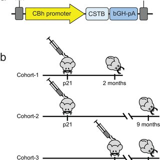
a AAV9 vector with a CBh promoter driving expression of human CSTB (AAV-CSTB). B AAV-CSTB or PBS administrated to three cohorts of Cstb−/− mice intrathecally. Cohort 1 and 2 were both injected at P21 (early time point) and brains examined at 2 and 9 months, respectively. Mice in cohort 3 were injected at p60 and brains examined at 9 months. Wild type (WT) littermates were left untreated in each cohort and used as controls.
Human CSTB protein expression is detected in the mouse brain
AAV9-CSTB and PBS-treated representative mouse brain sections (lower right insets) from a cohort 1, c cohort 2, and e cohort 3, were subjected to immunohistochemistry using a human specific CSTB antibody. Scale bar is 1 mm. Western blot analysis was performed using brain tissue from AAV9-CSTB and PBS-treated mice of cohorts 1(b), 2 (d), and 3 (f).
CSTB expression lowers expression of early-onset neuroinflammation markers
Relative mRNA expression levels of neuroinflammation markers Cxcl1, Cxcl10, Cxcl13, Iba1, and Gfap, were analyzed in cohorts 1 (a), 2 (b), and 3 (c) by qRT-PCR. Cohort 1, N = 15, per group. Cohort 2 and 3, WT (N = 13), PBS (N = 20), AAV-CSTB (N = 15). Data are presented as mean ± SEM. *p < 0.05, **p < 0.01, ***p < 0.001, and ****p < 0.0001. ns denotes non-significant.
AAV-CSTB gene replacement reduces neuronal apoptosis but does not prevent brain atrophy
The weight of ground brain tissue from one hemisphere of a. cohort 1 (p21) at the age of 2 months and b. cohort 2 (p21) and 3 (p60) at the age of 7–9 months, was measured. Data represent the means of WT (N = 14 for cohort 1 and N = 12 for cohort 2 & 3), PBS (N = 13 for cohort 1 and N = 20 for cohort 2 and 3), AAV-CSTB p21 (N = 13 for cohort 1 and N = 15 for cohort 2 and 3) ± SEM. c Apoptotic bodies were detected in cerebellar sections using TUNEL assays. Propidium iodide (PI) was used for counterstaining. d Quantitative analysis for the number of apoptotic bodies in cerebellar sections is shown. Scale bars are 1 mm. Data represent the mean of WT (N = 16), PBS (N = 11) and AAV-CSTB (N = 15) ± SEM. ****p < 0.0001. ns denotes non-significant.
The effect of AAV-CSTB gene therapy on the performance of Cstb−/− mice in the rotarod test
Cohort 2 (p21) and cohort 3 (p60) were subjected to both stationary and rotating (2 rpm) rotarods. Data represent the means of WT (N = 18), PBS (N = 21), and AAV-CSTB (N = 14) ± SEM for cohort 1, and WT (N = 18), PBS (N = 21) and AAV-CSTB (N = 15) ± SEM for cohort 2. *p < 0.05, **p < 0.01, ***p < 0.001, and ****p < 0.0001. ns denotes non-significant.
CSTB gene replacement improves neuroinflammation, neurodegeneration and ataxia in murine type 1 progressive myoclonus epilepsy

December 2023

·

53 Reads

·

1 Citation

Gene Therapy

EPM1 is the most common form of Progressive Myoclonus Epilepsy characterized by late-childhood onset, ever-worsening and disabling myoclonus, seizures, ataxia, psychiatric disease, and shortened lifespan. EPM1 is caused by expansions of a dodecamer repeat sequence in the promoter of CSTB (cystatin B), which dramatically reduces, but does not eliminate, gene expression. The relatively late onset and consistent presence of a minimal amount of protein product makes EPM1 a favorable target for gene replacement therapy. If treated early, these children’s normally developed brains could be rescued from the neurodegeneration that otherwise follows, and their cross-reactive immunological material (CRIM) positive status greatly reduces transgene related toxicity. We performed a proof-of-concept CSTB gene replacement study in Cstb knockout mice by introducing full-length human CSTB driven by the CBh promoter packaged in AAV9 and administered at postnatal days 21 and 60. Mice were sacrificed at 2 or 9 months of age, respectively. We observed significant improvements in expression levels of neuroinflammatory pathway genes and cerebellar granule cell layer apoptosis, as well as amelioration of motor impairment. The data suggest that gene replacement is a promising therapeutic modality for EPM1 and could spare affected children and families the ravages of this otherwise severe neurodegenerative disease.


Fig 1. Burden of variants for different CVS thresholds across epilepsy phenotypes. Odds ratios and p-value were calculated using a binomial logistic regression for variants of different Constraint Violation Score (CVS) thresholds. Lines represent 95% confidence intervals. Comparisons were made for cases and controls (A), Genetic Generalized Epilepsy (GGE) and controls (B), Non-Acquired Focal Epilepsy (NAFE) and controls (C) and GGE and NAFE (D). https://doi.org/10.1371/journal.pone.0291935.g001
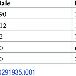
Number of individuals for each phenotype.
Unraveling the role of non-coding rare variants in epilepsy

September 2023

·

51 Reads

PLOS ONE

PLOS ONE

The discovery of new variants has leveled off in recent years in epilepsy studies, despite the use of very large cohorts. Consequently, most of the heritability is still unexplained. Rare non-coding variants have been largely ignored in studies on epilepsy, although non-coding single nucleotide variants can have a significant impact on gene expression. We had access to whole genome sequencing (WGS) from 247 epilepsy patients and 377 controls. To assess the functional impact of non-coding variants, ExPecto, a deep learning algorithm was used to predict expression change in brain tissues. We compared the burden of rare non-coding deleterious variants between cases and controls. Rare non-coding highly deleterious variants were significantly enriched in Genetic Generalized Epilepsy (GGE), but not in Non-Acquired Focal Epilepsy (NAFE) or all epilepsy cases when compared with controls. In this study we showed that rare non-coding deleterious variants are associated with epilepsy, specifically with GGE. Larger WGS epilepsy cohort will be needed to investigate those effects at a greater resolution. Nevertheless, we demonstrated the importance of studying non-coding regions in epilepsy, a disease where new discoveries are scarce.


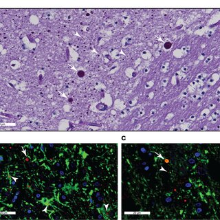
Figure 1. Lafora body (LB) Accumulation and Initial Home-Cage Response. A: Representative PAS-D images showing LB accumulation (TOP) and immunohistochemical assessments of LB accumulation (GS1), astrogliosis (GFAP) and microglial activation (Iba1). Scale bar: 50 µm. B: Home-cage structure and representative aerial video snapshot. C: Raster plot of distances traversed every minute of the 2-hour long introduction trial for every mouse, with measures of licking, shelter and feeder engagement. D: Heatmaps (left) and trackmaps (right) for a representative WT and KO mouse. Mean + s.e.m shown for all.
Figure 4. Changes in Home-cage Behavior with Age. A: On baseline day 2, compared with 6-week old mice, older WT cohorts displayed diminished sucrose preference (F2,56 = 2.94, p = 0.06) and feeding durations (F2,56 = 10.55, p<0,0001). B: Measures of sleep timing and duration were unchanged. C,D: 6-month and 1-year old mice displayed a blunted response to light-spot and BEEP stimulation. E: Older mice displayed fewer wheel rotations (F2,56 = 9.10, p<0.001). Mean + s.e.m shown for all. *, **, ***, **** depict p<0.05, <0.01, <0.001 or <0.0001 respectively.
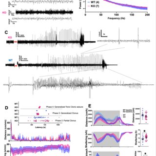
Figure 5. EEG and PTZ Responses. A: Representative single-channel electrocorticography from 1-year old WT and KO mice. B: EEG power spectra calculated during wakefulness. C: Representative EEG responses to a single intraperitoneal injection of PTZ (60mg/kg), demonstrating a prolonged epoch of spike/wave discharges, followed by a discrete epoch of evolving rhythmicity. Red bars annotate epochs of absent EEG signal while the mouse receives the intraperitoneal injection. D: Distance and sheltering responses to a single subconvulsant PTZ injection (30mg/kg), with a tally of convulsive events (inset) for both WT (n=14) and KO (n = 18). E: Post-ictal period home-cage metrics, revealing a comparative increase in sheltering and reduction in feeding in KO mice. Mean + s.e.m shown for all. * depicts p<0.05.
Figure 6: UP States in WT vs KO Somatosensory Cortex. A: Example traces of extracellular recordings in brain slices exhibiting spontaneously occurring activity bursts (from 12-month-old mice). B: The average duration of activity bursts in MKO slices is longer at 3 and 6 months of age, but not at 12 months (MannWhitney test, *p< 0.05) C: Burst amplitudes and frequencies. D: Relative power over all activity. Mean + s.e.m shown for all. Sample sizes at 3 months: WT (9 slices, 4 mice), KO (13 slices, 5 mice). 6 months: WT (19 slices, 6 mice), KO (29 slices, 7 mice). 12 months: WT (12 slices, 6 mice), KO (15 slices, 5 mice).
Figure 7: Lafora body accumulation in mouse piriform cortex (malin KO, 12 months of age).
Clinicopathologic Dissociation: Robust Lafora Body Accumulation in Malin KO Mice Without Observable Changes in Home-cage Behavior

September 2023

·

41 Reads

Lafora Disease (LD) is a syndrome of progressive myoclonic epilepsy and cumulative neurocognitive deterioration caused by recessively inherited genetic lesions of EPM2A (laforin) or NHLRC1 (malin). Neuropsychiatric symptomatology in LD is thought to be directly downstream of neuronal and astrocytic polyglucosan aggregates, termed Lafora bodies (LBs), which faithfully accumulate in an age-dependent manner in all mouse models of LD. In this study, we applied home-cage monitoring to examine the extent of neurobehavioral deterioration in a model of malin-deficient LD, as a means to identify robust preclinical endpoints that may guide the selection of novel genetic treatments. At 6 weeks, ~6-7 months and ~12 months of age, malin deficient mice (KO) and wild type (WT) littermates underwent a standardized home-cage behavioral assessment designed to non-obtrusively appraise features of rest/arousal, consumptive behaviors, risk aversion and voluntary wheel-running. At all timepoints, and over a range of metrics that we report transparently, WT and KO mice were essentially indistinguishable. In contrast, within WT mice compared across timepoints, we identified age-related nocturnal hypoactivity, diminished sucrose preference and reduced wheel-running. Neuropathological examinations in subsets of the same mice revealed expected age dependent LB accumulation, gliosis and microglial activation in cortical and subcortical brain regions. At 12 months of age, despite the burden of neocortical LBs, we did not identify spontaneous seizures during an electroencephalographic (EEG) survey, and KO and WT mice exhibited similar spectral EEG features. Using an in vitro assay of neocortical function, paroxysmal increases in network activity (UP states) in KO slices were more prolonged at 3 and 6 months of age, but were similar to WT at 12 months. KO mice displayed a distinct response to pentylenetetrazole, with a greater incidence of clonic seizures and a more pronounced post-ictal suppression of movement, feeding and drinking behavior. Together, these results highlight a stark clinicopathologic dissociation in a mouse model of LD, where LBs accrue substantially without clinically meaningful changes in overall wellbeing. Our findings allude to a delay between LB accumulation and neurobehavioral decline: one that may provide a window for treatment, and whose precise duration may be difficult to ascertain within the typical lifespan of a laboratory mouse.


Citations (58)


... We read with interest the article by Ozlu et al. about a retrospective, single-centre observational study of 50 patients diagnosed with a mitochondrial disorder (MID) collected over a 10-year period [1] . A pathogenic nDNA mutation was identified in 27 patients, a pathogenic mtDNA mutation in 17 patients, and no causative mutation in 6 patients. ...

Reference:

Assessing Genotype-phenotype Correlations in Mitochondrial Disorders Requires Genetic and Prospective Clinical Studies
Mitochondrial encephalopathies and myopathies: Our tertiary center's experience
  • Citing Article
  • March 2024

European Journal of Paediatric Neurology

... However, each PME has its unique features, as we have hopefully made clear throughout this review. Breakthroughs both in the pathophysiology and in the therapies for certain PMEs, such as LD, ULD, and MERRF are advancing rapidly, including a publication from our group showing evidence in favor of gene therapy in a murine model of ULD [149]. For other PMEs, progress has been more incremental. ...

CSTB gene replacement improves neuroinflammation, neurodegeneration and ataxia in murine type 1 progressive myoclonus epilepsy

Gene Therapy

... 21,22 Besides, previous studies have found that targeting the GYS1 could halt the progression of epilepsy and neuroinflammation. [23][24][25] Additionally, numerous studies have shown that GYS1 participates in tumor growth and progression through different mechanisms such as via the nuclear factor kappa-B (NF-κB) pathway, the AMP-activated protein kinase (AMPK) pathway, and the hypoxic inducible factor-1 alpha (HIF-1α) pathway. [26][27][28] The enzymatic function of GYS1 can be regulated through post-translational modification and the influence of allosteric effectors. ...

Gys1 Antisense Therapy Prevents Disease-Driving Aggregates and Epileptiform Discharges in a Lafora Disease Mouse Model

Journal of the American Society for Experimental NeuroTherapeutics

... Absences of the enzymes laforin or malin also result in amylopectinosis and neurodegeneration, in this case associated with a fatal teenage-onset progressive myoclonus epilepsy (Lafora disease, LD) [22,70]. Laforin is a glycogen phosphatase [108,125] as well as a scaffold protein for the localization of malin at glycogen [71]. Malin is an E3 ubiquitin ligase [41]. ...

Lafora progressive myoclonus-epilepsy: Laforin targets malin to glycogen

Disease Models and Mechanisms

... AAV gene replacement therapy can be used for the delivery of genes encoding neutrophic factors, enzymes, potassium channels, and neuromodulatory peptides, as examples [134]. A recent review discussing the genetics of SLC13A5 deficiency disorder, a DEE caused by deficiency of the sodium/citrate cotransporter, highlighted some logistical considerations for the development of AAV-based gene therapy which can be applied when considering gene therapy in the IESS population [135]. First, due to the size of AAV vectors, genes must be less than 5 kilobases in order to be packaged. ...

SLC13A5 Deficiency Disorder: From Genetics to Gene Therapy

Genes

... Furthermore, mouse models allow for the identification and validation of potential biomarkers associated with monogenic epilepsy, which can be instrumental in diagnosing the condition, predicting its progression, and monitoring an individual's response to treatment [40]. Lastly, mouse models provide a preclinical testing platform for evaluating novel therapies, such as gene therapy, targeted drugs, and neurostimulation techniques, before their translation into clinical trials [41]. These avenues highlight the significant role of mouse models in furthering our knowledge and improving therapeutic approaches in the field of monogenic epilepsy. ...

Gene Therapy: Novel Approaches to Targeting Monogenic Epilepsies

... The CENet cohort is composed of patients with Genetic Generalized Epilepsy (GGE) or Non-Acquired Focal Epilepsy (NAFE) collected in CHUM Research Center in Montreal and controls (unaffected Developmental Epileptic Encephalopathy (DEE) trio parents) collected in CHU Ste-Justine in Montreal and the Hospital for Sick Children in Toronto [23][24][25][26]. The patients were recruited between 2002 and 2014. ...

Assessment of burden and segregation profiles of CNVs in patients with epilepsy

... This occurs even in cases of monogenic epilepsy, such as Dravet syndrome (associated with SCN1A variants), developmental epileptic encephalopathies (DEE), and other forms of epilepsy associated with intellectual disability. 14,15 For this reason, using cell models derived from the patient is highly advantageous due to the preservation of the genetic background. However, when using patient-derived cultures, it is difficult to determine the contribution of individual gene variants to disease development. ...

The role of common genetic variation in presumed monogenic epilepsies

EBioMedicine

... Glycogen becomes polyglucosan when glycogen synthase outpaces GBE1 [15,62,68,69,85,122]. In animal models of all three amylopectinoses discussed here, glycogen no longer becomes polyglucosan and PBs are prevented from being formed when glycogen synthase is downregulated, whether by allelic knockout, RNA interference or small molecule enzyme inhibition [3,24,45,46,76,109,113]. The pathogenesis of polyglucosans in APBD is clear, namely branching enzyme deficiency, but remains unknown in LD and PGBM1. ...

AAV-Mediated Artificial miRNA Reduces Pathogenic Polyglucosan Bodies and Neuroinflammation in Adult Polyglucosan Body and Lafora Disease Mouse Models

Journal of the American Society for Experimental NeuroTherapeutics

... This was consistent with a prior finding of bipolar cell atrophy from Lafora body accumulation seen on histology. This finding was further confirmed in a subsequent study of six patients with LD-mild to severe generalized cone dysfunction of the retina was confirmed on ERG in all patients [51]. Interestingly, cone and rod dysfunction correlated with both the disease duration and the type of mutation. ...

Ocular phenotype and electroretinogram abnormalities in Lafora disease and correlation with disease stage

Journal of Neurology